Copyright
©The Author(s) 2023.
World J Clin Cases. Mar 6, 2023; 11(7): 1569-1575
Published online Mar 6, 2023. doi: 10.12998/wjcc.v11.i7.1569
Published online Mar 6, 2023. doi: 10.12998/wjcc.v11.i7.1569
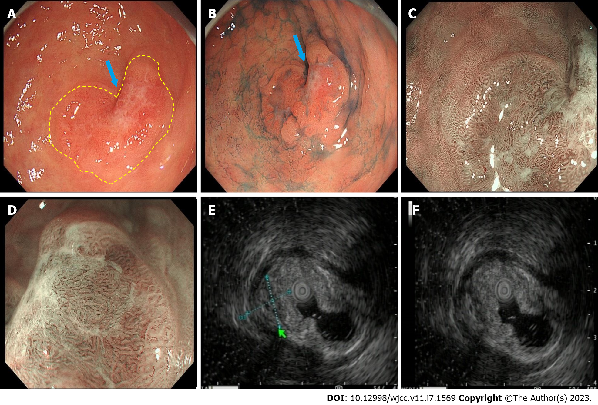
Figure 1 The major gastric lesion.
A: White light imaging demonstrating a heart-shaped lesion. The demarcation line is highlighted with a yellow dashed line. The arrow indicates the ectopic pancreatic opening; B: Indigo carmine dying; C and D: Narrow band imaging combined with magnifying endoscopy; E and F: Endoscopic ultrasound demonstrating hypoechoic changes, uneven internal echoes and unclear boundaries between some areas and muscularis propria in the major lesion.
- Citation: Zhao ZY, Lai YX, Xu P. Gastric ectopic pancreas combined with synchronous multiple early gastric cancer: A rare case report. World J Clin Cases 2023; 11(7): 1569-1575
- URL: https://www.wjgnet.com/2307-8960/full/v11/i7/1569.htm
- DOI: https://dx.doi.org/10.12998/wjcc.v11.i7.1569


