Copyright
©The Author(s) 2023.
World J Clin Cases. Mar 6, 2023; 11(7): 1560-1568
Published online Mar 6, 2023. doi: 10.12998/wjcc.v11.i7.1560
Published online Mar 6, 2023. doi: 10.12998/wjcc.v11.i7.1560
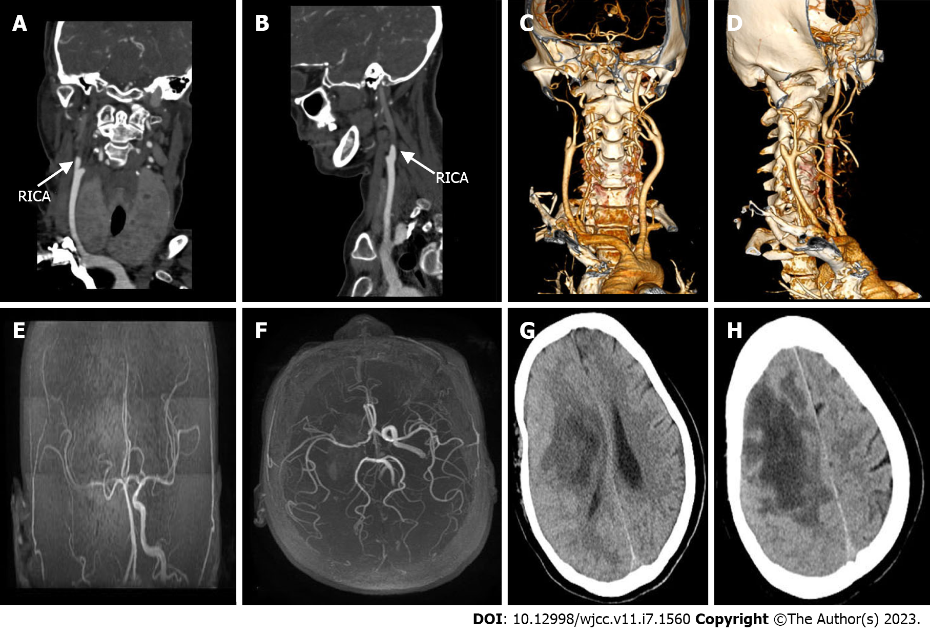
Figure 2 Vascular examination and head computed tomography review.
A-D: The right internal carotid artery was occluded, and the distal end was not shown. No obvious abnormalities were observed in the left carotid artery and vertebral artery; E and F: The right internal carotid artery skull base and intracranial segment were occluded, and the anterior and posterior cerebral arteries were supplied by the traffic branch; G and H: The right frontal lobe and basal ganglia showed lamellar low-density shadows, most of which tended to be liquid density, the right lateral ventricle was slightly stressed, and the midline was shifted to the left. RICA: Right internal carotid artery.
- Citation: Chen CH, Chen JN, Du HG, Guo DL. Isolated cerebral mucormycosis that looks like stroke and brain abscess: A case report and review of the literature. World J Clin Cases 2023; 11(7): 1560-1568
- URL: https://www.wjgnet.com/2307-8960/full/v11/i7/1560.htm
- DOI: https://dx.doi.org/10.12998/wjcc.v11.i7.1560


