Copyright
©The Author(s) 2023.
World J Clin Cases. Mar 6, 2023; 11(7): 1560-1568
Published online Mar 6, 2023. doi: 10.12998/wjcc.v11.i7.1560
Published online Mar 6, 2023. doi: 10.12998/wjcc.v11.i7.1560
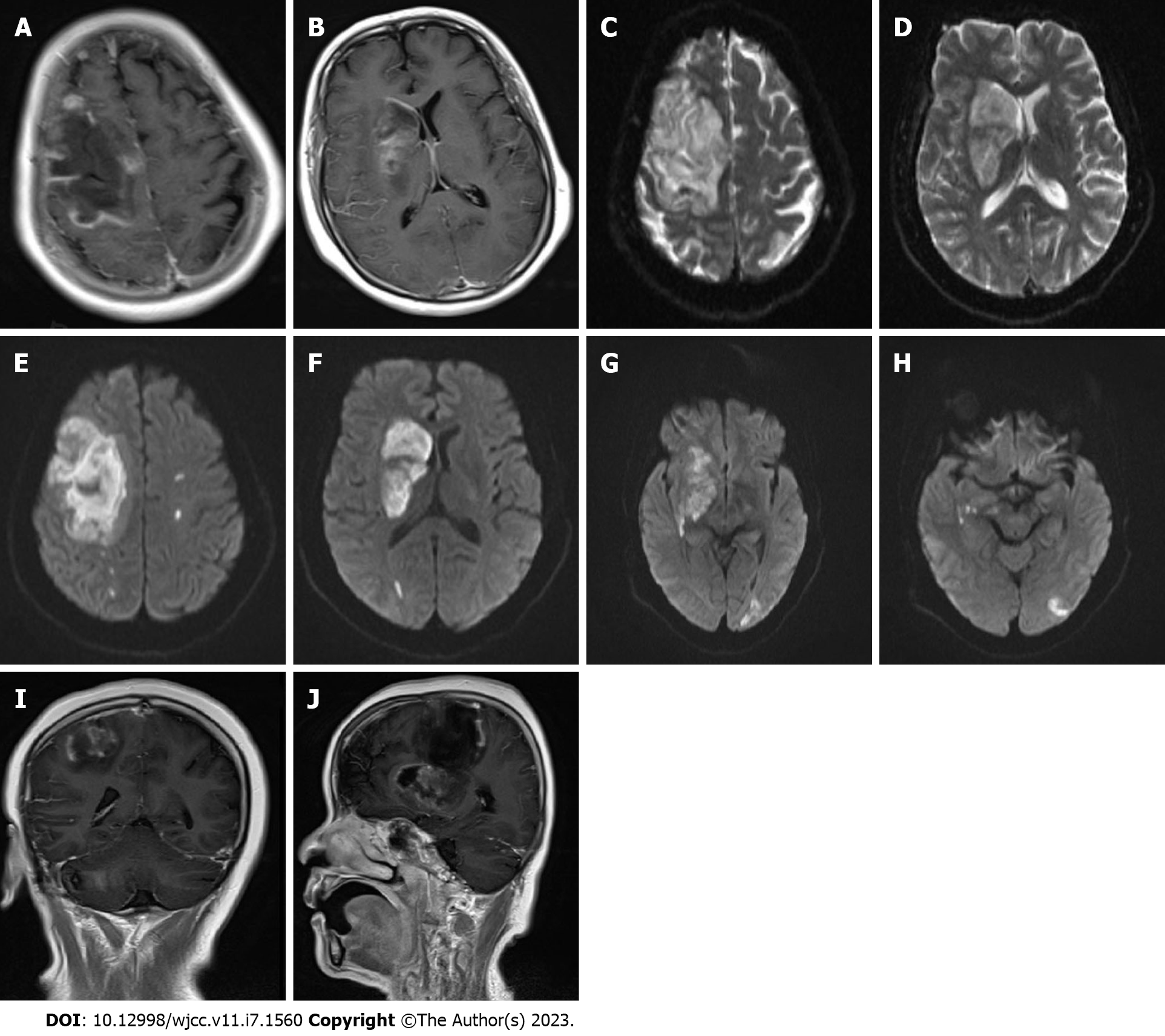
Figure 1 Head magnetic resonance imaging findings.
A-D: In the right frontal lobes and basal ganglia, large-scale abnormal signal lesions were found with low signal on T1-weighted imaging and slightly higher signal on T2-weighted imaging; E-H: Diffusion-weighted imaging signals were enhanced in the right frontal lobe and right basal ganglia, and multiple spotted high signals were observed in the left hemisphere; I and J: After enhancement, no ring enhancement was observed around the lesion, and there was no obvious space-occupying effect.
- Citation: Chen CH, Chen JN, Du HG, Guo DL. Isolated cerebral mucormycosis that looks like stroke and brain abscess: A case report and review of the literature. World J Clin Cases 2023; 11(7): 1560-1568
- URL: https://www.wjgnet.com/2307-8960/full/v11/i7/1560.htm
- DOI: https://dx.doi.org/10.12998/wjcc.v11.i7.1560


