Copyright
©The Author(s) 2023.
World J Clin Cases. Mar 6, 2023; 11(7): 1528-1548
Published online Mar 6, 2023. doi: 10.12998/wjcc.v11.i7.1528
Published online Mar 6, 2023. doi: 10.12998/wjcc.v11.i7.1528
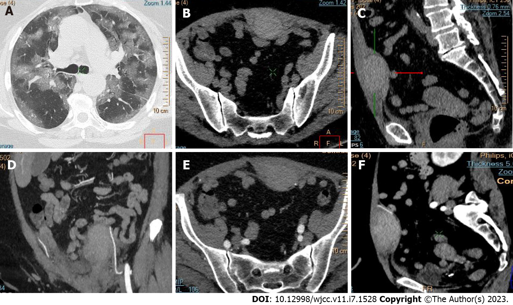
Figure 12 Thoracic and pelvic computed tomography images of hematoma of the anterior abdominal wall.
A: Diffuse “ground glass” opacifications from coronavirus disease 2019 bilateral pneumonia; B and C: Pre-contrast axial and coronal images; D-F: Coronal, axial, and sagittal reconstructions with intravenous contrast show a mild hematoma in the left rectus abdominis muscle due to bleeding from the left inferior epigastric artery.
- Citation: Evrev D, Sekulovski M, Gulinac M, Dobrev H, Velikova T, Hadjidekov G. Retroperitoneal and abdominal bleeding in anticoagulated COVID-19 hospitalized patients: Case series and brief literature review. World J Clin Cases 2023; 11(7): 1528-1548
- URL: https://www.wjgnet.com/2307-8960/full/v11/i7/1528.htm
- DOI: https://dx.doi.org/10.12998/wjcc.v11.i7.1528


