Copyright
©The Author(s) 2023.
World J Clin Cases. Mar 6, 2023; 11(7): 1528-1548
Published online Mar 6, 2023. doi: 10.12998/wjcc.v11.i7.1528
Published online Mar 6, 2023. doi: 10.12998/wjcc.v11.i7.1528
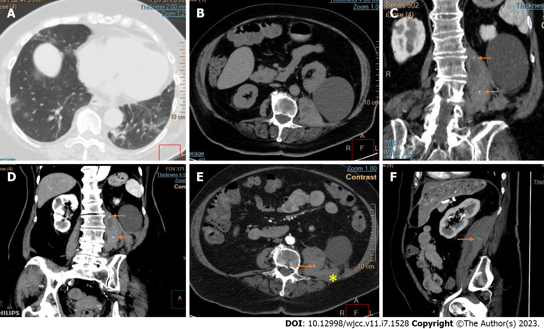
Figure 8 Abdominal pre- and post-contrast images and thoracic computed tomography in a patient with coronavirus disease 2019 and hematoma of the left psoas muscle.
A: “Crazy caving” pattern and linear interstitial thickening in the basal lung segments in organizing coronavirus disease 2019 pneumonia; B-F: A large hematoma of the left psoas muscle with infiltration of quadratus lumborum (asterisk); B-E: Large simple cortical cyst of the left kidney; C-F: Active bleeding from lumbar branches of the iliolumbar artery is indicated on post-contrast computed tomography images (arrows).
- Citation: Evrev D, Sekulovski M, Gulinac M, Dobrev H, Velikova T, Hadjidekov G. Retroperitoneal and abdominal bleeding in anticoagulated COVID-19 hospitalized patients: Case series and brief literature review. World J Clin Cases 2023; 11(7): 1528-1548
- URL: https://www.wjgnet.com/2307-8960/full/v11/i7/1528.htm
- DOI: https://dx.doi.org/10.12998/wjcc.v11.i7.1528


