Copyright
©The Author(s) 2023.
World J Clin Cases. Mar 6, 2023; 11(7): 1528-1548
Published online Mar 6, 2023. doi: 10.12998/wjcc.v11.i7.1528
Published online Mar 6, 2023. doi: 10.12998/wjcc.v11.i7.1528
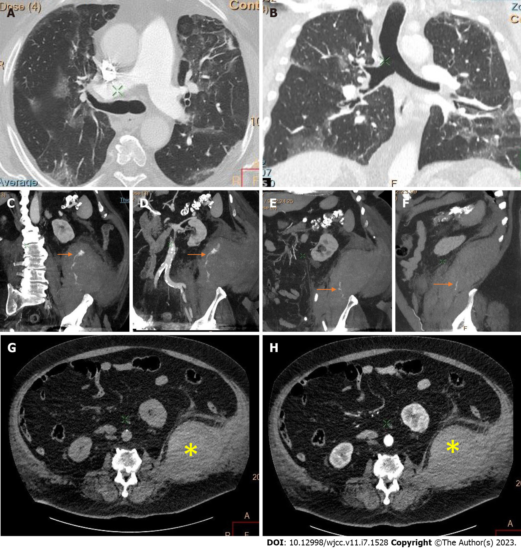
Figure 3 Thoracic and abdominal computed tomography of a large retroperitoneal hematoma.
A: Bilateral lung involvement with coronavirus disease 2019 (COVID-19) pneumonia in cross section; and B: In vertical section; C-F: Multiplanar reconstructions present the site of active bleeding and the jet from the extravasated contrast material (arrows); C-H: The hematoma arises from the internal, the external oblique, and the transverse abdominal muscles and invades the adjacent psoas, quadratus lumborum, and iliacus muscles on the left, which appear thickened (asterisk).
- Citation: Evrev D, Sekulovski M, Gulinac M, Dobrev H, Velikova T, Hadjidekov G. Retroperitoneal and abdominal bleeding in anticoagulated COVID-19 hospitalized patients: Case series and brief literature review. World J Clin Cases 2023; 11(7): 1528-1548
- URL: https://www.wjgnet.com/2307-8960/full/v11/i7/1528.htm
- DOI: https://dx.doi.org/10.12998/wjcc.v11.i7.1528


