Copyright
©The Author(s) 2023.
World J Clin Cases. Mar 6, 2023; 11(7): 1521-1527
Published online Mar 6, 2023. doi: 10.12998/wjcc.v11.i7.1521
Published online Mar 6, 2023. doi: 10.12998/wjcc.v11.i7.1521
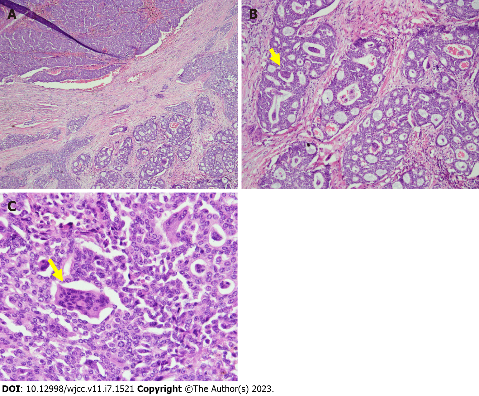
Figure 3 Breast Histopathology.
A: Low-power view of the infiltrative mammary tumor, composed of ducts, nests, and cribriform patterns; B: Medium-power view demonstrates osteoclast-like stromal giant cells (yellow arrow) and red blood cell extravasation; C: High-power view, multinucleated OGCs (yellow arrow) embedded in invasive breast carcinoma.
- Citation: Wang YJ, Huang CP, Hong ZJ, Liao GS, Yu JC. Invasive breast carcinoma with osteoclast-like stromal giant cells: A case report. World J Clin Cases 2023; 11(7): 1521-1527
- URL: https://www.wjgnet.com/2307-8960/full/v11/i7/1521.htm
- DOI: https://dx.doi.org/10.12998/wjcc.v11.i7.1521


