Copyright
©The Author(s) 2023.
World J Clin Cases. Mar 6, 2023; 11(7): 1498-1505
Published online Mar 6, 2023. doi: 10.12998/wjcc.v11.i7.1498
Published online Mar 6, 2023. doi: 10.12998/wjcc.v11.i7.1498
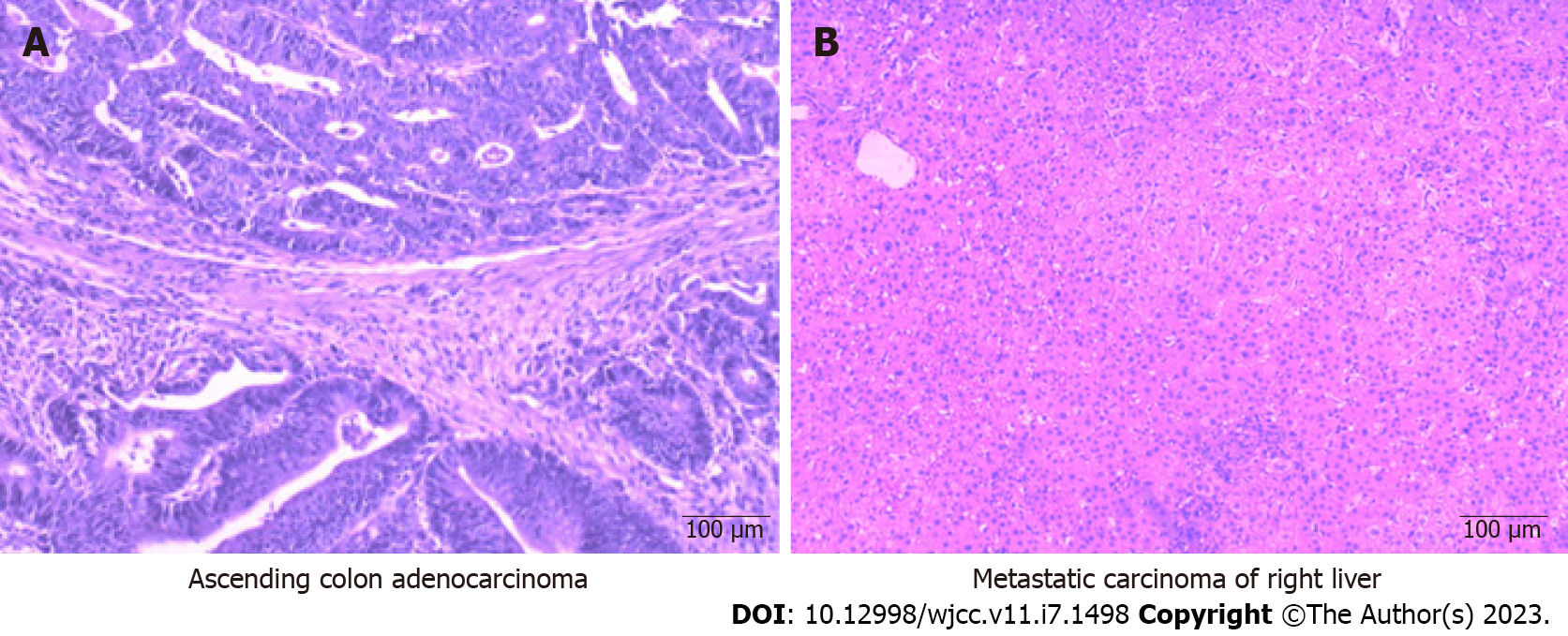
Figure 2 Hematoxylin-eosin staining (× 100).
A: The resected specimen (right colon) showed a massive moderately differentiated adenocarcinoma with necrosis. The cancer tissue invaded the entire intestinal wall and involved the nerves. Intratumoral vascular thrombosis was observed, and no cancer metastasis was seen in the peri-intestinal lymph nodes; B: The surgical resection specimen (right liver tumor) showed no cancerous cells in the liver metastasis, fibrous tissue hyperplasia, collagenous necrosis, and inflammatory cell infiltration, surrounding liver tissue congestion, hemorrhage, or inflammatory cell infiltration.
- Citation: Tan XR, Li J, Chen HW, Luo W, Jiang N, Wang ZB, Wang S. Successful multidisciplinary therapy for a patient with liver metastasis from ascending colon adenocarcinoma: A case report and review of literature. World J Clin Cases 2023; 11(7): 1498-1505
- URL: https://www.wjgnet.com/2307-8960/full/v11/i7/1498.htm
- DOI: https://dx.doi.org/10.12998/wjcc.v11.i7.1498


