Copyright
©The Author(s) 2023.
World J Clin Cases. Mar 6, 2023; 11(7): 1498-1505
Published online Mar 6, 2023. doi: 10.12998/wjcc.v11.i7.1498
Published online Mar 6, 2023. doi: 10.12998/wjcc.v11.i7.1498
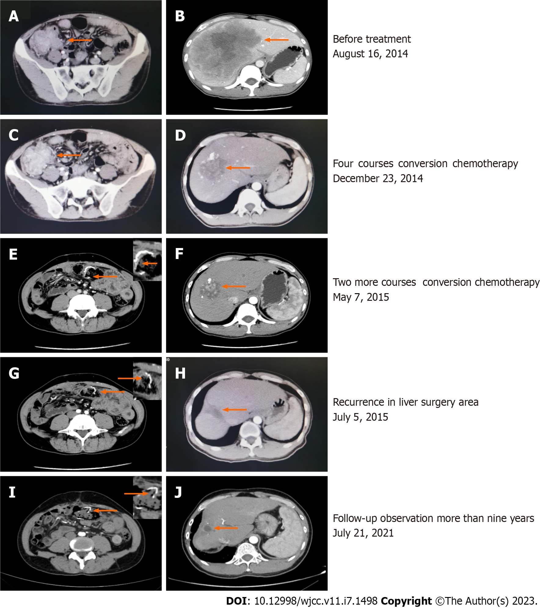
Figure 1 Enhanced computed tomography.
A: Malignant tumors in the ileocecal area and lymph nodes of varying sizes were observed adjacent to the mesangium, and metastasis was suspected; B: A huge liver metastasis in the right lobe of the liver, approximately 13.7 cm × 14.1 cm in size (arrow), with compression of the right portal vein; C and D: After four courses of treatment, the tumor in the ascending colon was not significantly reduced (6.8 to 5.4 cm), and the low-density metastatic lesion in the liver had shrunk from 14.1 to 5.9 cm; E, H, and I: The anastomosis in the colon cancer surgery area was unobstructed, and no abnormally enhanced lesions were seen; F: After another 2 cycles of conversion chemotherapy, the liver mass was significantly reduced to approximately 5.2 cm in size, with multiple lipiodol deposits on the edges; G: Residual liver parenchymal nodular enhancement in the right lobe of the liver (3.0 cm in length) that was considered a postoperative recurrence; J: A sheet-like low-density shadow was seen in the right lobe of the liver, with a size of approximately 1.6 cm × 1.5 cm, and no abnormal enhancement was observed.
- Citation: Tan XR, Li J, Chen HW, Luo W, Jiang N, Wang ZB, Wang S. Successful multidisciplinary therapy for a patient with liver metastasis from ascending colon adenocarcinoma: A case report and review of literature. World J Clin Cases 2023; 11(7): 1498-1505
- URL: https://www.wjgnet.com/2307-8960/full/v11/i7/1498.htm
- DOI: https://dx.doi.org/10.12998/wjcc.v11.i7.1498


