Copyright
©The Author(s) 2023.
World J Clin Cases. Feb 26, 2023; 11(6): 1419-1425
Published online Feb 26, 2023. doi: 10.12998/wjcc.v11.i6.1419
Published online Feb 26, 2023. doi: 10.12998/wjcc.v11.i6.1419
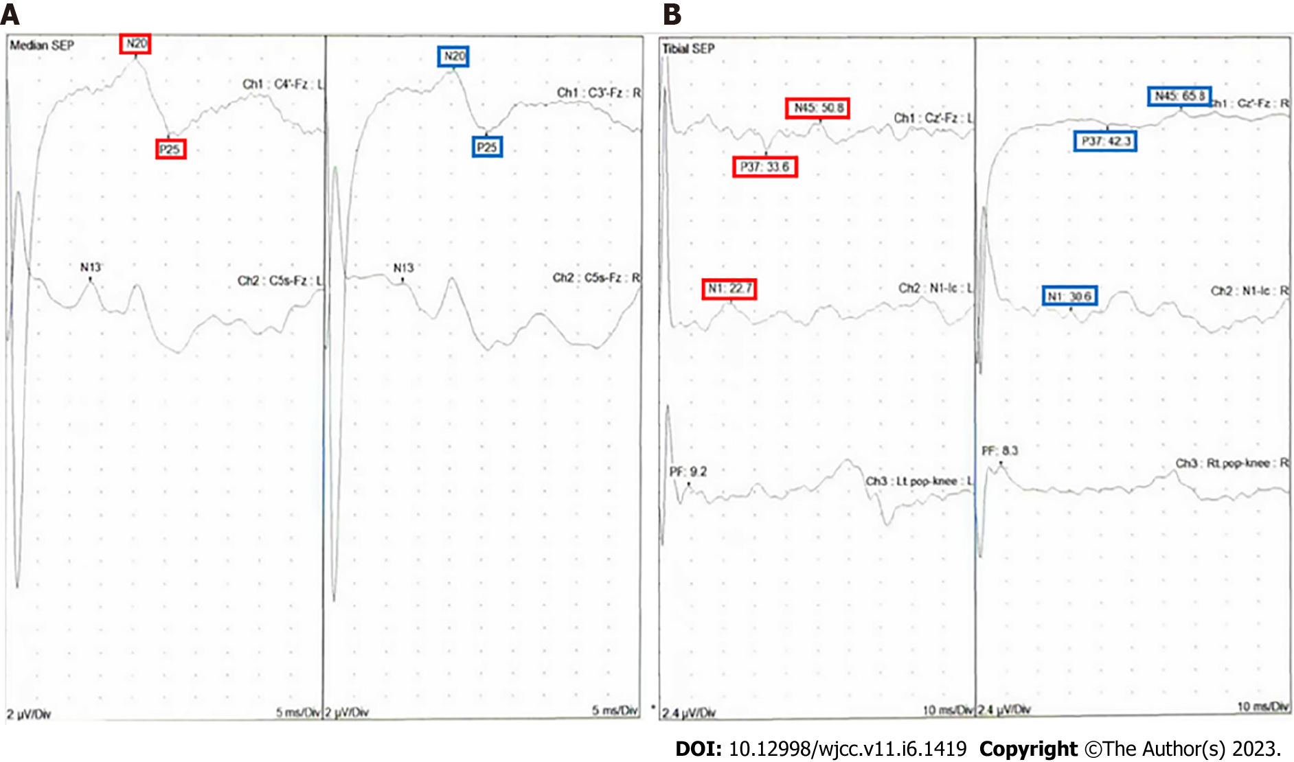
Figure 3 The somatosensory evoked potentials were recorded by alternately stimulating each posterior tibial nerve at the ankle region behind the medial malleolus or the median nerve at the wrist.
Two averages of 120 trials were obtained to stimulate each nerve. The somatosensory evoked potentials revealed well-developed cortical peaks for either arm. A: The principal peaks of N20 and P25 were 17 and 21 ms for both MNs; B: The peaks of both P37, N45, and N1 were delayed with the right specific in posterior tibial nerve. No side-to-side latency difference was noted.
- Citation: Cho SY, Jang BH, Seo JW, Kim SW, Lim KJ, Lee HY, Kim DJ. Transverse myelitis caused by herpes zoster following COVID-19 vaccination: A case report. World J Clin Cases 2023; 11(6): 1419-1425
- URL: https://www.wjgnet.com/2307-8960/full/v11/i6/1419.htm
- DOI: https://dx.doi.org/10.12998/wjcc.v11.i6.1419


