Copyright
©The Author(s) 2023.
World J Clin Cases. Feb 26, 2023; 11(6): 1356-1364
Published online Feb 26, 2023. doi: 10.12998/wjcc.v11.i6.1356
Published online Feb 26, 2023. doi: 10.12998/wjcc.v11.i6.1356
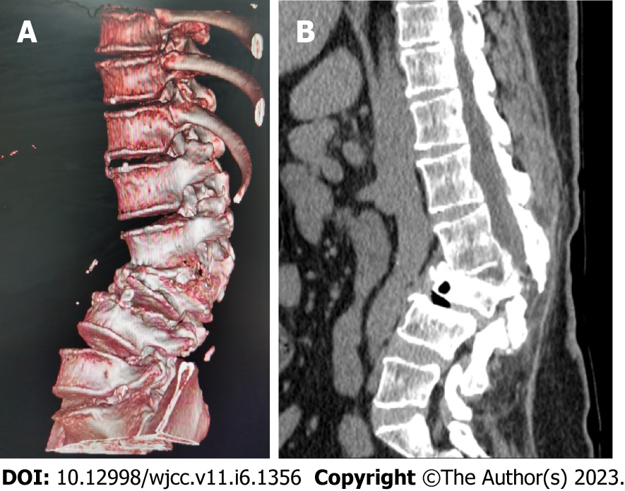
Figure 7 Lumbar vertebral body (plain scan + 3D reconstruction) computed tomography.
Lumbar vertebral body (plain scan + 3D reconstruction) computed tomography (CT) showed: L2-3 plane lumbar lordosis, L2-3 vertebral space narrowing, L3 vertebral body wedge-shaped flattening, L3, 4 vertebral body adjacent edge patchy high density. There are some bone defects in the lamina and spinous process of L3 and L4 vertebrae, and the vertebral canal at the same plane is deformed. A: Lumbar vertebral body CT (3D reconstruction); B: Lumbar vertebral body CT (plain scan).
- Citation: Liu YD, Deng Q, Li JJ, Yang HY, Han XF, Zhang KD, Peng RD, Xiang QQ. Post-traumatic cauda equina nerve calcification: A case report. World J Clin Cases 2023; 11(6): 1356-1364
- URL: https://www.wjgnet.com/2307-8960/full/v11/i6/1356.htm
- DOI: https://dx.doi.org/10.12998/wjcc.v11.i6.1356


