Copyright
©The Author(s) 2023.
World J Clin Cases. Feb 26, 2023; 11(6): 1356-1364
Published online Feb 26, 2023. doi: 10.12998/wjcc.v11.i6.1356
Published online Feb 26, 2023. doi: 10.12998/wjcc.v11.i6.1356
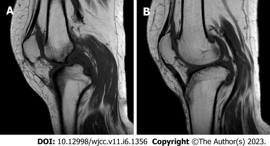
Figure 6 The right knee magnetic resonance imaging.
The cartilage of the medial femoral condyle and tibial plateau is worn, the cartilage is denatured, the posterior cruciate ligament of the right knee is torn, the medial collateral ligament is injured, the anterior and posterior corners of the medial and lateral meniscus of the right knee are worn, the right knee joint has effusion, and the medial head bursa of the gastrocnemius has effusion. A: Lateral magnetic resonance imaging (MRI) of knee; B: Oblique MRI of knee.
- Citation: Liu YD, Deng Q, Li JJ, Yang HY, Han XF, Zhang KD, Peng RD, Xiang QQ. Post-traumatic cauda equina nerve calcification: A case report. World J Clin Cases 2023; 11(6): 1356-1364
- URL: https://www.wjgnet.com/2307-8960/full/v11/i6/1356.htm
- DOI: https://dx.doi.org/10.12998/wjcc.v11.i6.1356


