Copyright
©The Author(s) 2023.
World J Clin Cases. Feb 16, 2023; 11(5): 1217-1223
Published online Feb 16, 2023. doi: 10.12998/wjcc.v11.i5.1217
Published online Feb 16, 2023. doi: 10.12998/wjcc.v11.i5.1217
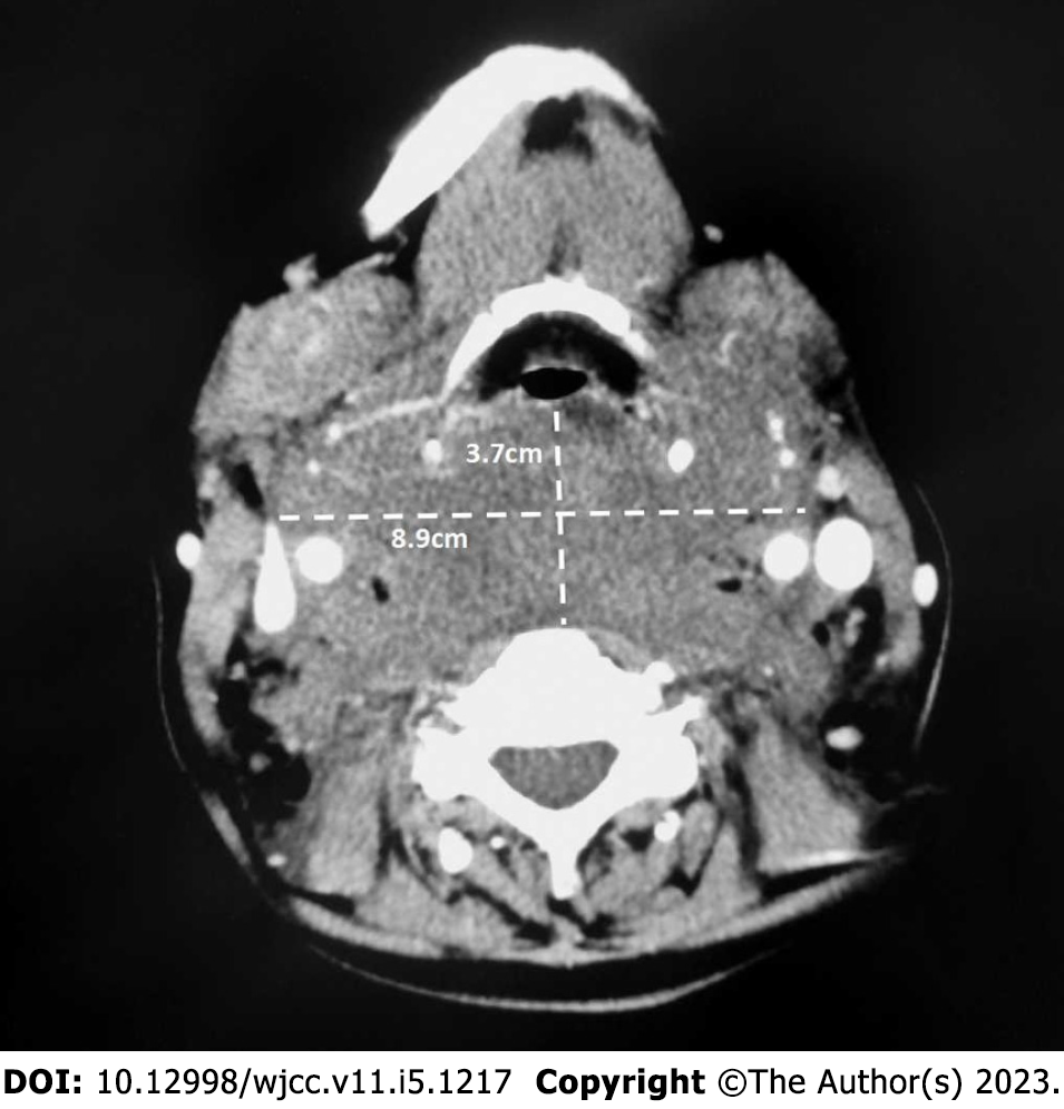
Figure 1 Preoperative axial venous phase contrast-enhanced computed tomography image of the neck (hyoid level).
The soft tissue density lesion is in contact with the posterior tracheal wall anteriorly, anterior cervical vertebra posteriorly, and the bilateral carotid sheaths, and is displacing the surrounding tissue and enveloping part of the vessels. There is no obvious enhancement of the lesion, which has a slightly lower density than muscle. There is swelling and effusion in the surrounding fatty tissue.
- Citation: Han YZ, Zhou Y, Peng Y, Zeng J, Zhao YQ, Gao XR, Zeng H, Guo XY, Li ZQ. Difficult airway due to cervical haemorrhage caused by spontaneous rupture of a parathyroid adenoma: A case report. World J Clin Cases 2023; 11(5): 1217-1223
- URL: https://www.wjgnet.com/2307-8960/full/v11/i5/1217.htm
- DOI: https://dx.doi.org/10.12998/wjcc.v11.i5.1217


