Copyright
©The Author(s) 2023.
World J Clin Cases. Feb 16, 2023; 11(5): 1188-1197
Published online Feb 16, 2023. doi: 10.12998/wjcc.v11.i5.1188
Published online Feb 16, 2023. doi: 10.12998/wjcc.v11.i5.1188
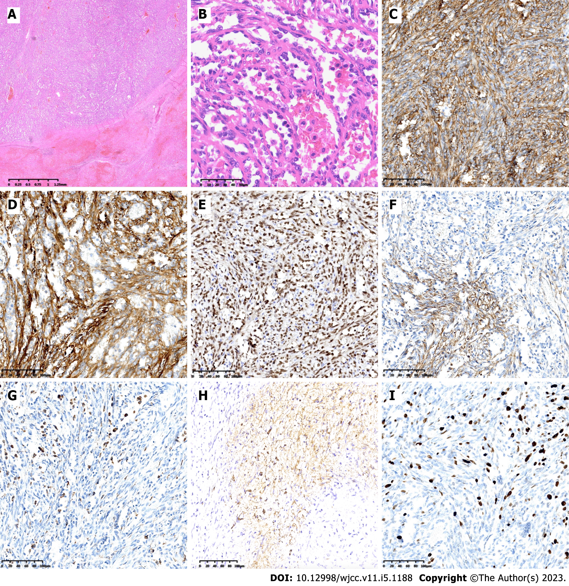
Figure 3 Pathological examination.
A: The lesion was located in the splenic red pulp, nearly all the surrounding tissues were invaded by the tumor cells (H&E magnification × 20); B: The lesion consists of a large amount of variably sized sinuses with anastomosing vascular channels lined by relatively plump round to columnar histiocytic-endothelial littoral cells, some cells with small nucleus with deep chromatin, some columnar cells with large nucleus with vacuolated chromatin, some exfoliated into the expanded sinuses (H&E magnification × 400); C-F: Immunohistochemical staining confirming endothelial differentiation with CD31, CD34, ERG, and FVIII (magnification × 200); G and H: Immunohistochemical staining confirming histiocytic differentiation with CD68 and CD21 (magnification × 200); I: Ki-67 labeling index was no more than 20% (magnification × 200).
- Citation: Jia F, Lin H, Li YL, Zhang JL, Tang L, Lu PT, Wang YQ, Cui YF, Yang XH, Lu ZY. Early postsurgical lethal outcome due to splenic littoral cell angioma: A case report. World J Clin Cases 2023; 11(5): 1188-1197
- URL: https://www.wjgnet.com/2307-8960/full/v11/i5/1188.htm
- DOI: https://dx.doi.org/10.12998/wjcc.v11.i5.1188


