Copyright
©The Author(s) 2023.
World J Clin Cases. Feb 16, 2023; 11(5): 1094-1098
Published online Feb 16, 2023. doi: 10.12998/wjcc.v11.i5.1094
Published online Feb 16, 2023. doi: 10.12998/wjcc.v11.i5.1094
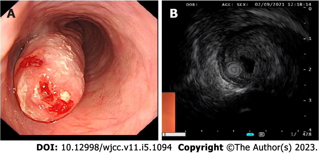
Figure 1 Imaging examinations.
A: Gastroscopy showed that a giant mass was located 30 cm from the incisor and extended to the cardia; B: Endoscopic ultrasonography showed that the mass was located in the submucosa, had mixed echo change, and exhibited some inner cystoid structures.
- Citation: Wang XS, Zhao CG, Wang HM, Wang XY. Giant myxofibrosarcoma of the esophagus treated by endoscopic submucosal dissection: A case report. World J Clin Cases 2023; 11(5): 1094-1098
- URL: https://www.wjgnet.com/2307-8960/full/v11/i5/1094.htm
- DOI: https://dx.doi.org/10.12998/wjcc.v11.i5.1094


