Copyright
©The Author(s) 2023.
World J Clin Cases. Feb 16, 2023; 11(5): 1086-1093
Published online Feb 16, 2023. doi: 10.12998/wjcc.v11.i5.1086
Published online Feb 16, 2023. doi: 10.12998/wjcc.v11.i5.1086
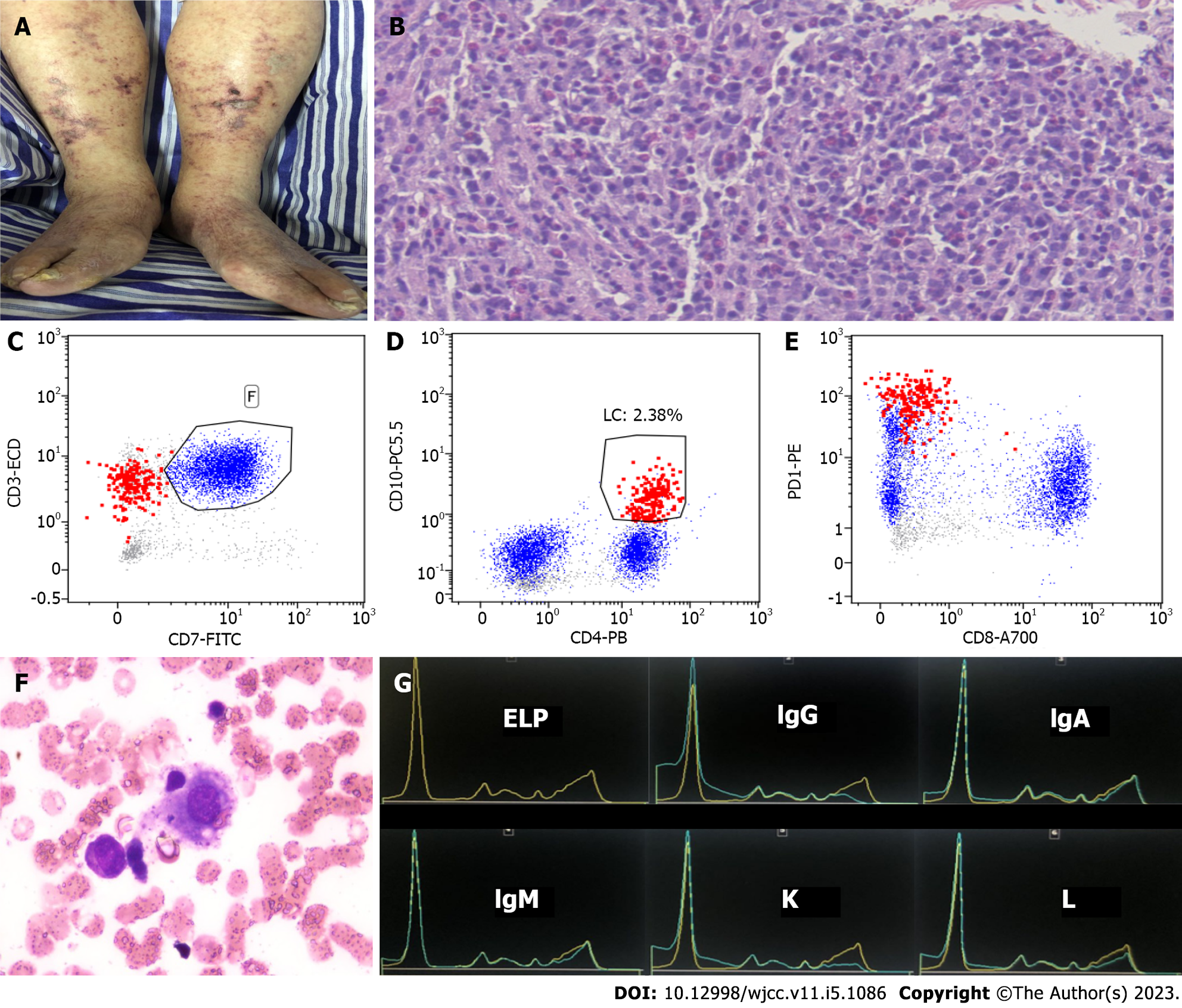
Figure 1 Examinations.
A: Purpura was observed on both lower limbs of the patient; B: Groin lymph node puncture specimen showed that the normal structure of lymph nodes disappeared and heterogeneous infiltration of small to medium-sized lymphoma cells, with proliferation of eosinophils (hematoxylin and eosin staining, × 40); C-E: Flow cytometry. Neoplastic T cells are shown in red and benign T cells in blue (analysis was gating on lymphocytes). The neoplastic T cells were positive for CD3, CD4, CD10, and PD1, but negative for CD7 and CD8; F: Bone marrow examination showed hemophagocytosis; G: Capillary electrophoresis revealed monoclonal IgG kappa.
- Citation: Jiang M, Wan JH, Tu Y, Shen Y, Kong FC, Zhang ZL. Angioimmunoblastic T-cell lymphoma induced hemophagocytic lymphohistiocytosis and disseminated intravascular coagulopathy: A case report. World J Clin Cases 2023; 11(5): 1086-1093
- URL: https://www.wjgnet.com/2307-8960/full/v11/i5/1086.htm
- DOI: https://dx.doi.org/10.12998/wjcc.v11.i5.1086


