Copyright
©The Author(s) 2023.
World J Clin Cases. Feb 6, 2023; 11(4): 962-971
Published online Feb 6, 2023. doi: 10.12998/wjcc.v11.i4.962
Published online Feb 6, 2023. doi: 10.12998/wjcc.v11.i4.962
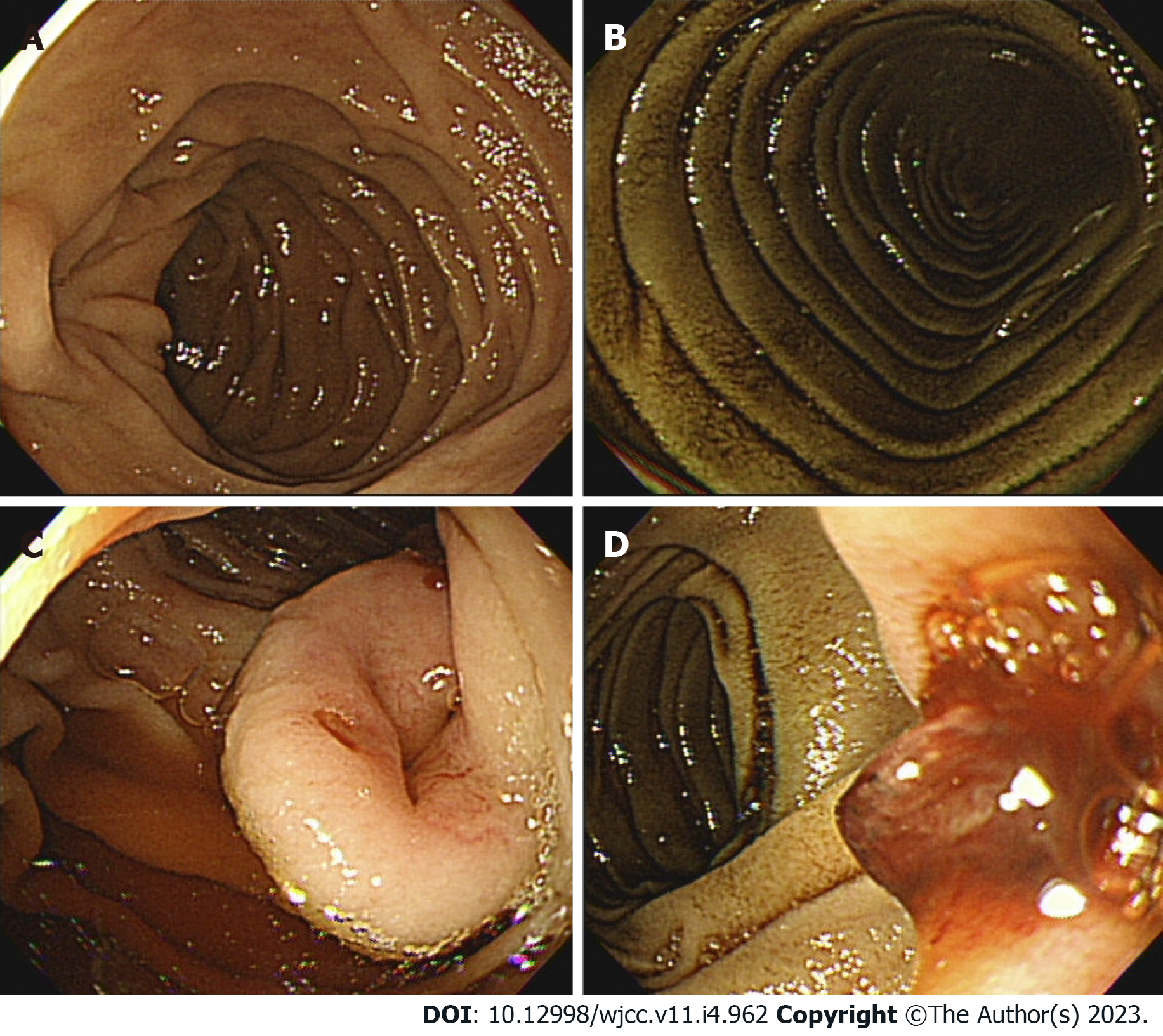
Figure 1 Conventional upper gastrointestinal endoscopy findings.
A and B: A dark discoloration is seen in the area which suspected to the third part of the duodenum; C: There is an approximately 2.5 cm central depressed mass in the transitional zone with mucosal color; D: The mass shows a smooth surface and focal bleeding with exposed vessels.
- Citation: Lee J, Kim S, Kim D, Lee S, Ryu K. Three cases of jejunal tumors detected by standard upper gastrointestinal endoscopy: A case series. World J Clin Cases 2023; 11(4): 962-971
- URL: https://www.wjgnet.com/2307-8960/full/v11/i4/962.htm
- DOI: https://dx.doi.org/10.12998/wjcc.v11.i4.962


