Copyright
©The Author(s) 2023.
World J Clin Cases. Feb 6, 2023; 11(4): 931-937
Published online Feb 6, 2023. doi: 10.12998/wjcc.v11.i4.931
Published online Feb 6, 2023. doi: 10.12998/wjcc.v11.i4.931
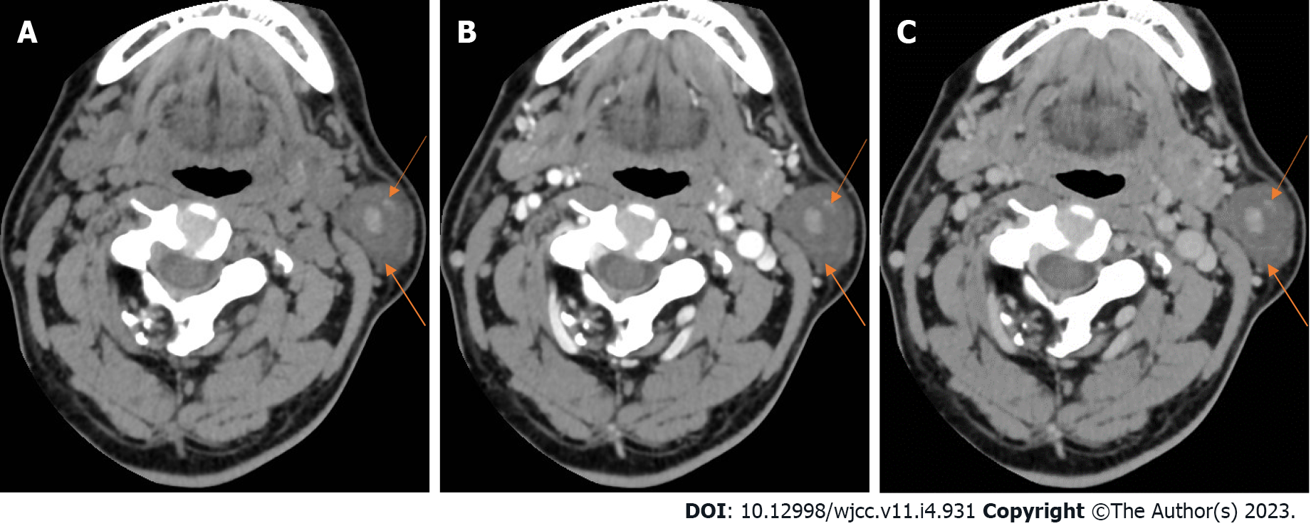
Figure 2 Neck computed tomography.
A: Plain computed tomography scan. A round cystic lesion was seen in the left parotid gland, with a maximum cross-sectional area of about 2.3 cm × 2.2 cm, uneven density, small patches with slightly high-density shadow, and clear boundary; B: Arterial phase. On enhanced scanning, the cyst wall was slightly enhanced in the arterial phase, but no obvious enhancement was observed in the cyst; C: Venous phase. The enhancement degree in the venous phase was similar to that in the arterial phase (orange arrows).
- Citation: Liao Y, Li YJ, Hu XW, Wen R, Wang P. Benign lymphoepithelial cyst of parotid gland without human immunodeficiency virus infection: A case report . World J Clin Cases 2023; 11(4): 931-937
- URL: https://www.wjgnet.com/2307-8960/full/v11/i4/931.htm
- DOI: https://dx.doi.org/10.12998/wjcc.v11.i4.931


