Copyright
©The Author(s) 2023.
World J Clin Cases. Feb 6, 2023; 11(4): 922-930
Published online Feb 6, 2023. doi: 10.12998/wjcc.v11.i4.922
Published online Feb 6, 2023. doi: 10.12998/wjcc.v11.i4.922
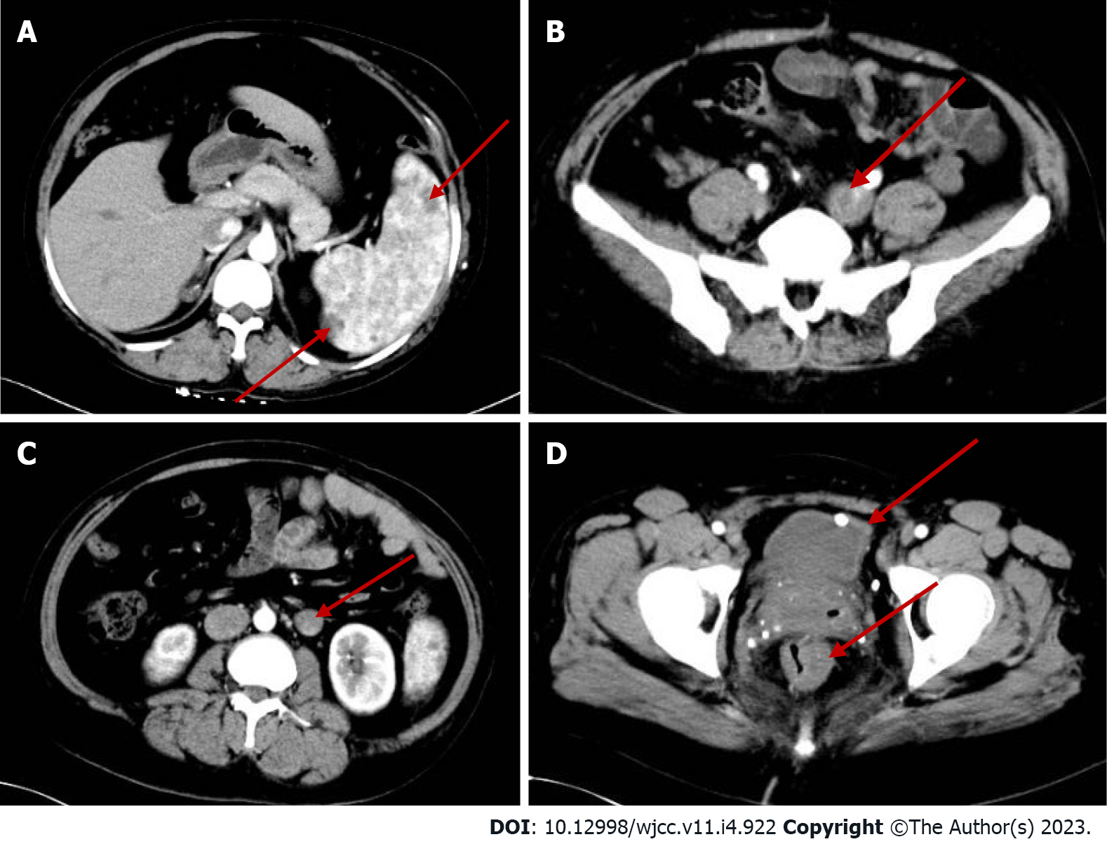
Figure 2 Abdominal computed tomography plain scan + enhancement.
A: The spleen is large with multiple low-density shadows (hemangioma); B: The left iliac vein is thickened; C: The left ovarian vein is thickened with a filling defect area (thrombosis); D: The rectal wall is unevenly thickened. The bladder wall is slightly thickened and venous stones can be observed.
- Citation: Li LL, Xie R, Li FQ, Huang C, Tuo BG, Wu HC. Easily misdiagnosed complex Klippel-Trenaunay syndrome: A case report. World J Clin Cases 2023; 11(4): 922-930
- URL: https://www.wjgnet.com/2307-8960/full/v11/i4/922.htm
- DOI: https://dx.doi.org/10.12998/wjcc.v11.i4.922


