Copyright
©The Author(s) 2023.
World J Clin Cases. Feb 6, 2023; 11(4): 725-737
Published online Feb 6, 2023. doi: 10.12998/wjcc.v11.i4.725
Published online Feb 6, 2023. doi: 10.12998/wjcc.v11.i4.725
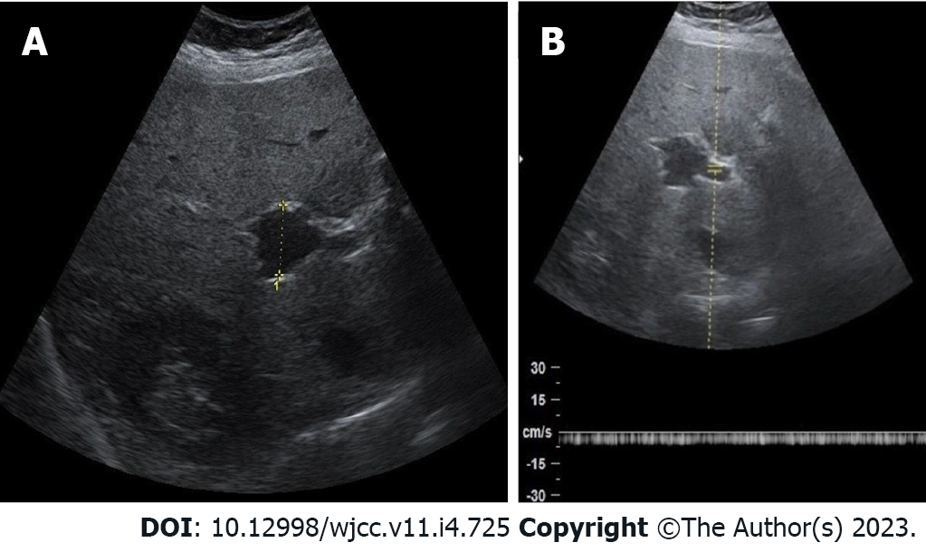
Figure 2 Sonography assessment of portal vein aneurysm.
A: Abdominal ultrasound shows portal vein aneurysm (PVA) at the level of bifurcation; B: Spectral Doppler sonography shows nonpulsatile blood flow through the portal venous system with PVA.
- Citation: Kurtcehajic A, Zerem E, Alibegovic E, Kunosic S, Hujdurovic A, Fejzic JA. Portal vein aneurysm-etiology, multimodal imaging and current management. World J Clin Cases 2023; 11(4): 725-737
- URL: https://www.wjgnet.com/2307-8960/full/v11/i4/725.htm
- DOI: https://dx.doi.org/10.12998/wjcc.v11.i4.725


