Copyright
©The Author(s) 2023.
World J Clin Cases. Dec 26, 2023; 11(36): 8527-8534
Published online Dec 26, 2023. doi: 10.12998/wjcc.v11.i36.8527
Published online Dec 26, 2023. doi: 10.12998/wjcc.v11.i36.8527
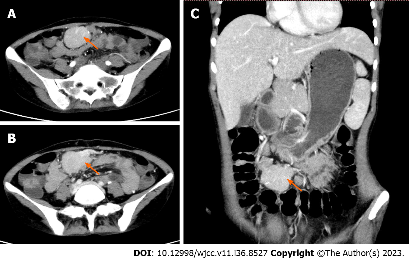
Figure 2 A mass of approximately 50 mm × 38 mm × 36 mm can be seen in the pelvic cavity in Case 2.
The boundary is clear and the shape is irregular. Small nodular calcification can be seen around the focus, and moderate enhancement was seen on the enhanced scan. A: Arterial phase; B: Venous phase; C: Coronal imaging.
- Citation: Gao JW, Shi ZY, Zhu ZB, Xu XR, Chen W. Intraperitoneal hyaline vascular Castleman disease: Three case reports. World J Clin Cases 2023; 11(36): 8527-8534
- URL: https://www.wjgnet.com/2307-8960/full/v11/i36/8527.htm
- DOI: https://dx.doi.org/10.12998/wjcc.v11.i36.8527


