Copyright
©The Author(s) 2023.
World J Clin Cases. Dec 16, 2023; 11(35): 8416-8424
Published online Dec 16, 2023. doi: 10.12998/wjcc.v11.i35.8416
Published online Dec 16, 2023. doi: 10.12998/wjcc.v11.i35.8416
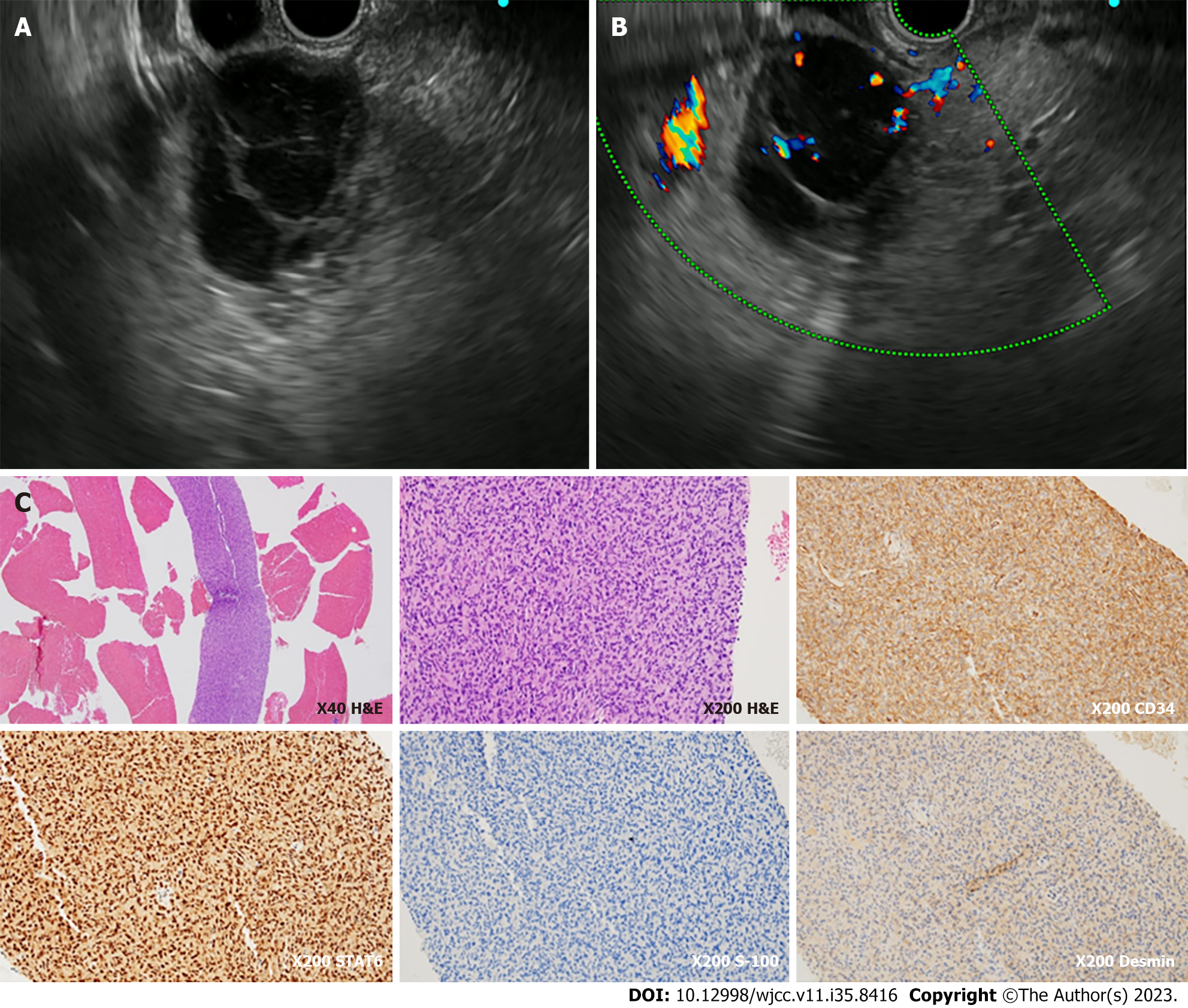
Figure 3 Endoscopic ultrasonography findings.
A: The mass showed hypoechogenicity inside, leading to an impression of cystic change; B: On doppler mode, blood vessels were observed inside the mass, suggesting it is less likely to be a cystic lesion; C: Endoscopic ultrasonography (EUS)-FNA specimen. H&E staining of the EUS-FNA specimen showed proliferation of spindle to ovoid cells. The specimen stained positive for smooth muscle actin, STAT6, and CD34, but negative for DOG-1, C-kit, S-100, and desmin.
- Citation: Yi K, Lee J, Kim DU. Metastatic pancreatic solitary fibrous tumor: A case report. World J Clin Cases 2023; 11(35): 8416-8424
- URL: https://www.wjgnet.com/2307-8960/full/v11/i35/8416.htm
- DOI: https://dx.doi.org/10.12998/wjcc.v11.i35.8416


