Copyright
©The Author(s) 2023.
World J Clin Cases. Dec 16, 2023; 11(35): 8416-8424
Published online Dec 16, 2023. doi: 10.12998/wjcc.v11.i35.8416
Published online Dec 16, 2023. doi: 10.12998/wjcc.v11.i35.8416
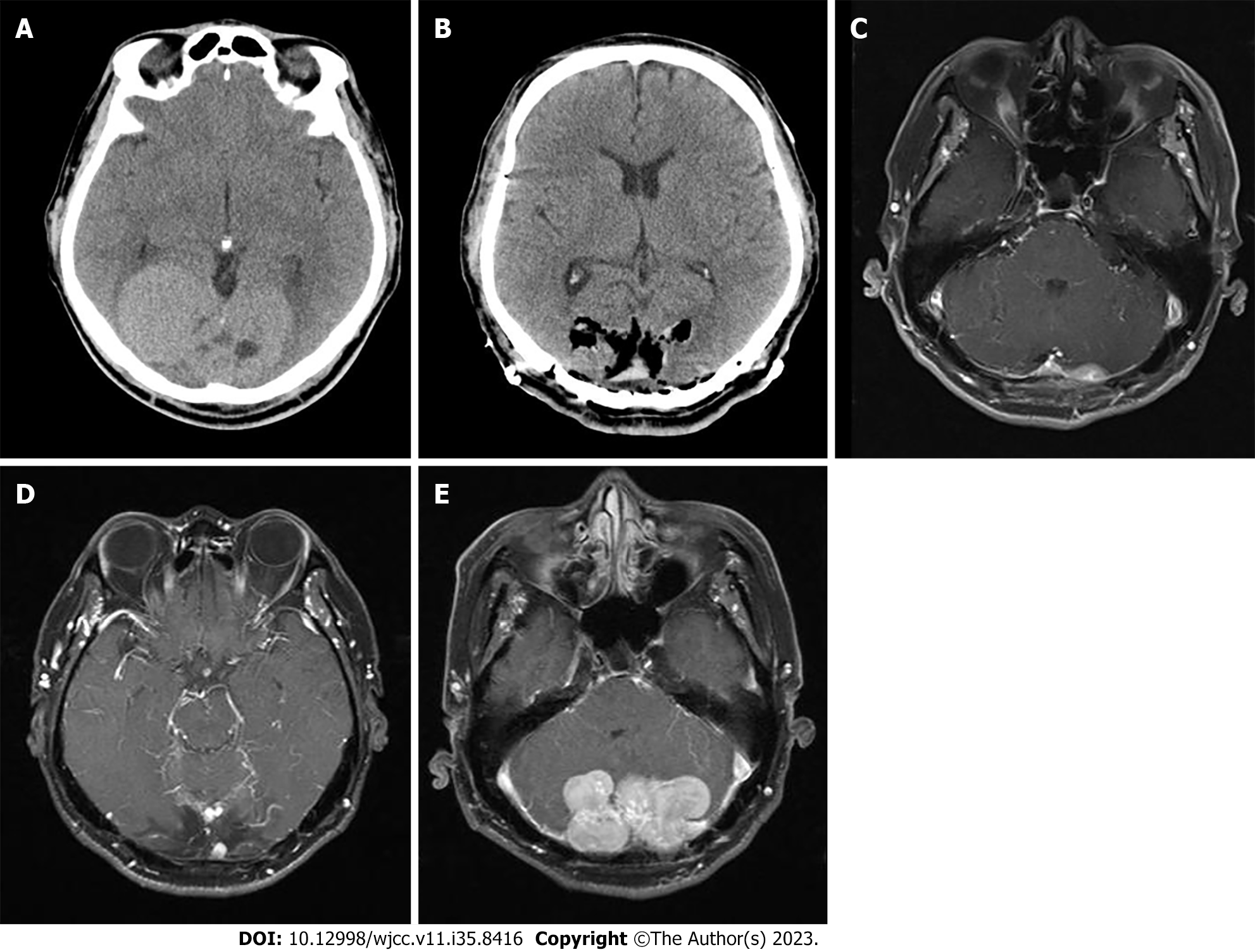
Figure 1 Time axis of the patient’s detection, treatment, and post-operative follow-up of brain tumor.
A: Brain computed tomography (CT) revealed an 8 cm × 4.7 cm multilobulated heterogeneous mass at both parieto-occipital lobe; B: Brain CT after osteoplastic craniotomy for removal; C: 16 mo later, brain magnetic resonance imaging (MRI) showed 1.7 cm enhancing lesion at the left cerebellar area which suggested remnant mass; D: 2 years later, brain MRI showed new 0.6 cm sized enhancing mass, suggesting recurrence; E: 5 years later, brain MRI showed multiple masses in both cerebellar area.
- Citation: Yi K, Lee J, Kim DU. Metastatic pancreatic solitary fibrous tumor: A case report. World J Clin Cases 2023; 11(35): 8416-8424
- URL: https://www.wjgnet.com/2307-8960/full/v11/i35/8416.htm
- DOI: https://dx.doi.org/10.12998/wjcc.v11.i35.8416


