Copyright
©The Author(s) 2023.
World J Clin Cases. Dec 16, 2023; 11(35): 8399-8403
Published online Dec 16, 2023. doi: 10.12998/wjcc.v11.i35.8399
Published online Dec 16, 2023. doi: 10.12998/wjcc.v11.i35.8399
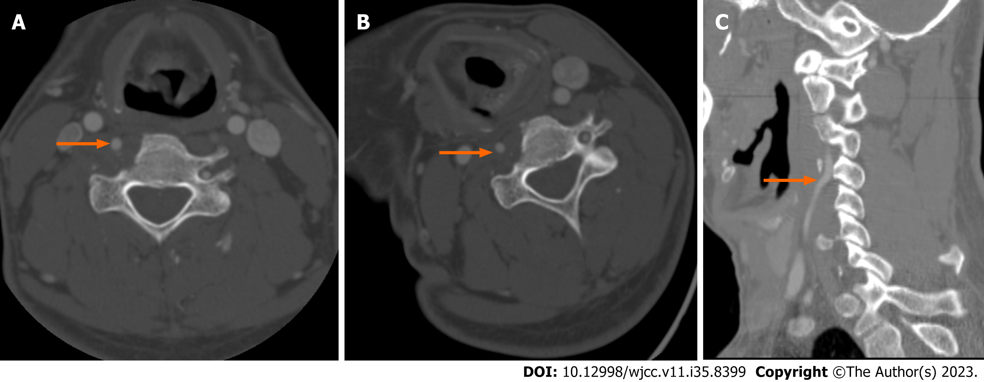
Figure 2 Postsurgical computed tomography angiography.
A: Anterior tubercle of C5 transverse process eliminated and right vertebral artery (VA) patent (arrow); B and C: Axial and sagittal images during right rotation of head showing no dynamic VA compression (arrows).
- Citation: Ahn JH, Jun HS, Kim IK, Kim CH, Lee SJ. Atypical case of bow hunter’s syndrome linked to aberrantly coursing vertebral artery: A case report. World J Clin Cases 2023; 11(35): 8399-8403
- URL: https://www.wjgnet.com/2307-8960/full/v11/i35/8399.htm
- DOI: https://dx.doi.org/10.12998/wjcc.v11.i35.8399


