Copyright
©The Author(s) 2023.
World J Clin Cases. Dec 16, 2023; 11(35): 8399-8403
Published online Dec 16, 2023. doi: 10.12998/wjcc.v11.i35.8399
Published online Dec 16, 2023. doi: 10.12998/wjcc.v11.i35.8399
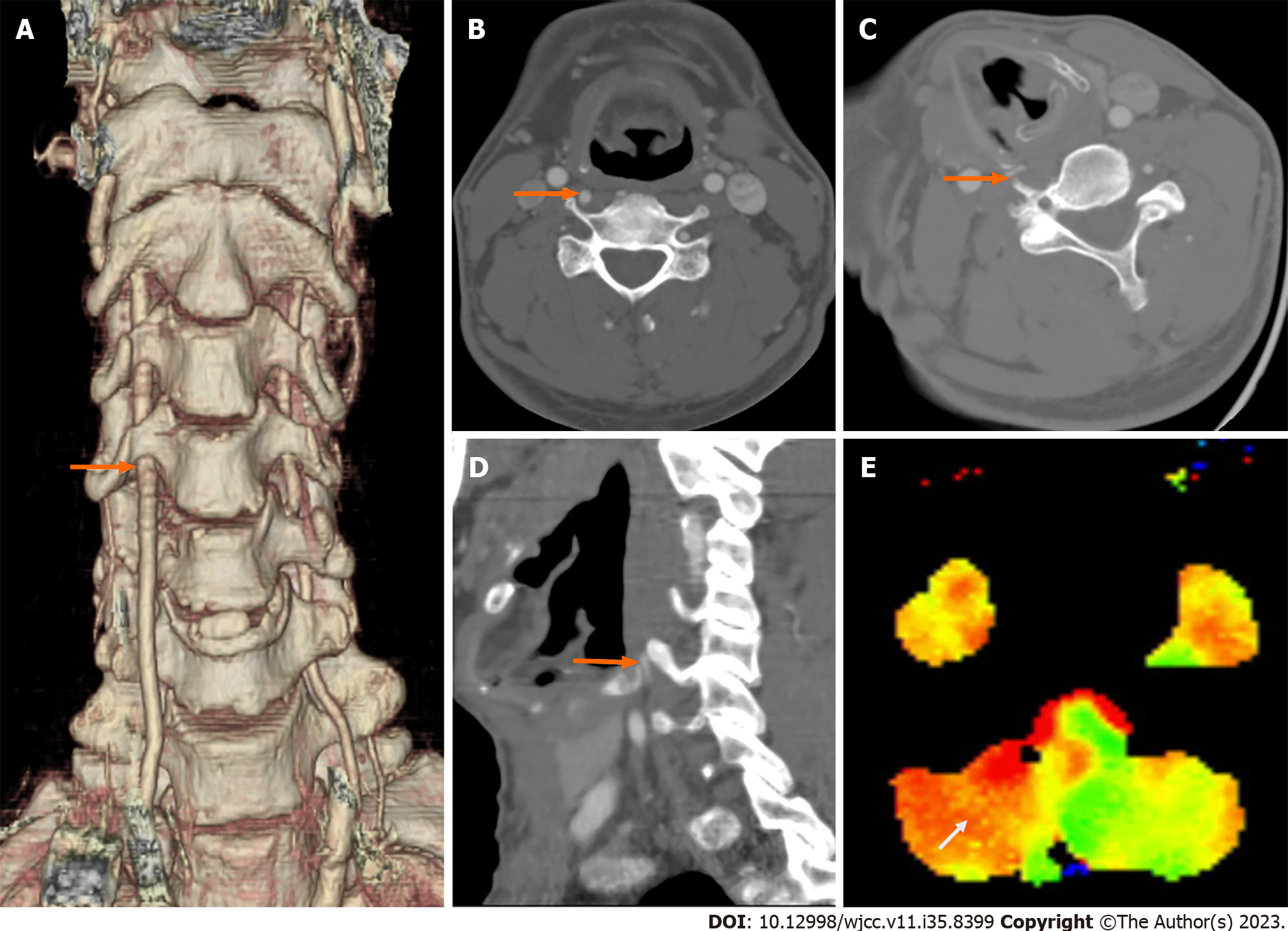
Figure 1 Preoperative computed tomography angiography and perfusion magnetic resonance imaging studies.
A: Normal C6 vertebral artery (VA) entry on left and anomalous right VA entry at C4 transverse foramen (arrow); B: Axial view of patent right VA in neutral position (arrow); C and D: Rotational dynamic compression of right VA by thyroid cartilage and anterior tubercle of C5 transverse process (arrows); E: Time-to-peak delay in right cerebellar territory (arrow) during head rotation to right.
- Citation: Ahn JH, Jun HS, Kim IK, Kim CH, Lee SJ. Atypical case of bow hunter’s syndrome linked to aberrantly coursing vertebral artery: A case report. World J Clin Cases 2023; 11(35): 8399-8403
- URL: https://www.wjgnet.com/2307-8960/full/v11/i35/8399.htm
- DOI: https://dx.doi.org/10.12998/wjcc.v11.i35.8399


