Copyright
©The Author(s) 2023.
World J Clin Cases. Dec 16, 2023; 11(35): 8392-8398
Published online Dec 16, 2023. doi: 10.12998/wjcc.v11.i35.8392
Published online Dec 16, 2023. doi: 10.12998/wjcc.v11.i35.8392
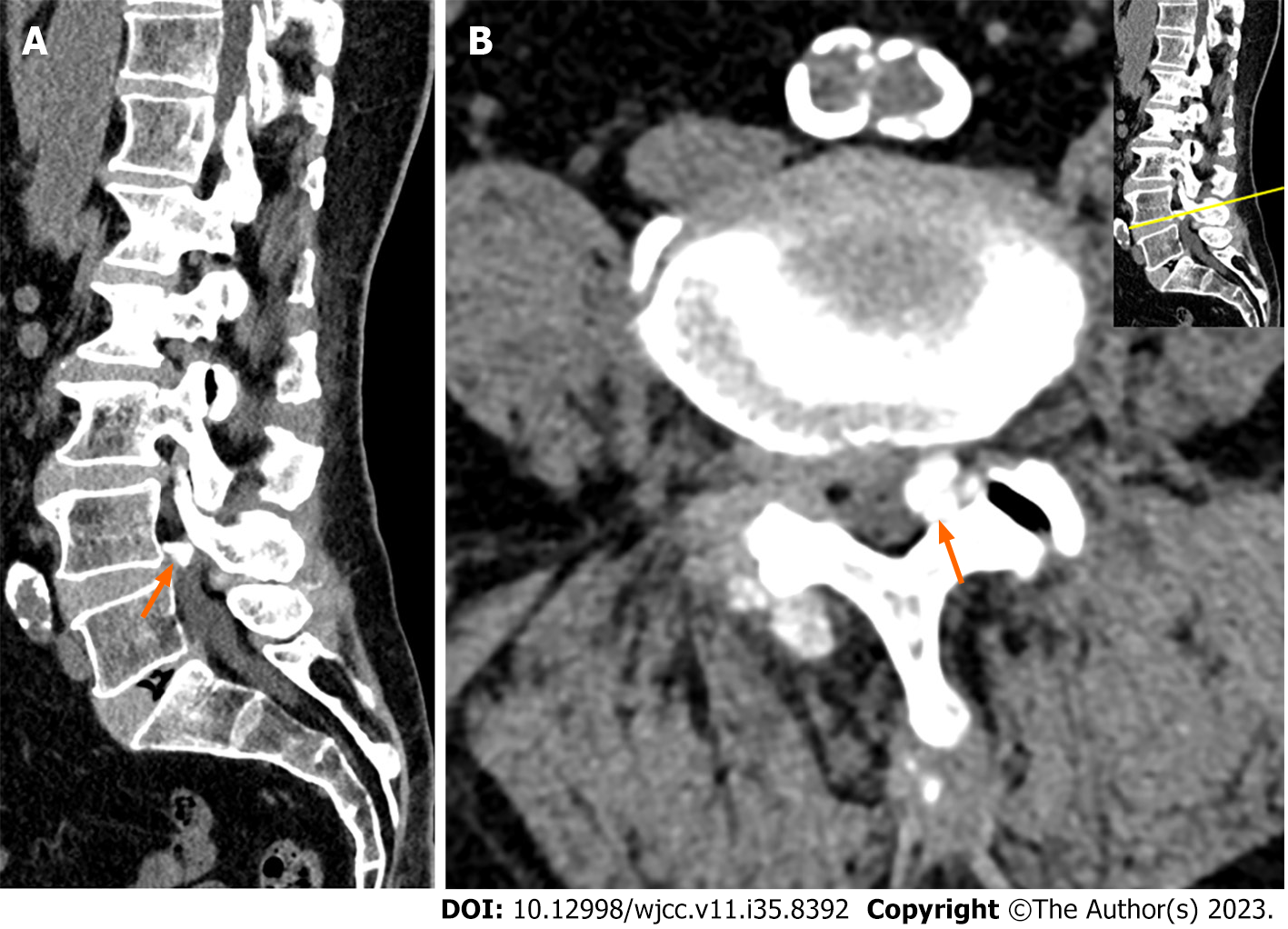
Figure 3 Computed tomography scans of the lumbar spine.
A: Sagittal computed tomography (CT) scan showing a calcified mass at the L4-5 level (arrow); B: Axial CT scan showing a calcified mass at the left side of the L4-5 level (arrow).
- Citation: Jung HY, Kim GU, Joh YW, Lee JS. Ankle and toe weakness caused by calcified ligamentum flavum cyst: A case report. World J Clin Cases 2023; 11(35): 8392-8398
- URL: https://www.wjgnet.com/2307-8960/full/v11/i35/8392.htm
- DOI: https://dx.doi.org/10.12998/wjcc.v11.i35.8392


