Copyright
©The Author(s) 2023.
World J Clin Cases. Dec 16, 2023; 11(35): 8343-8349
Published online Dec 16, 2023. doi: 10.12998/wjcc.v11.i35.8343
Published online Dec 16, 2023. doi: 10.12998/wjcc.v11.i35.8343
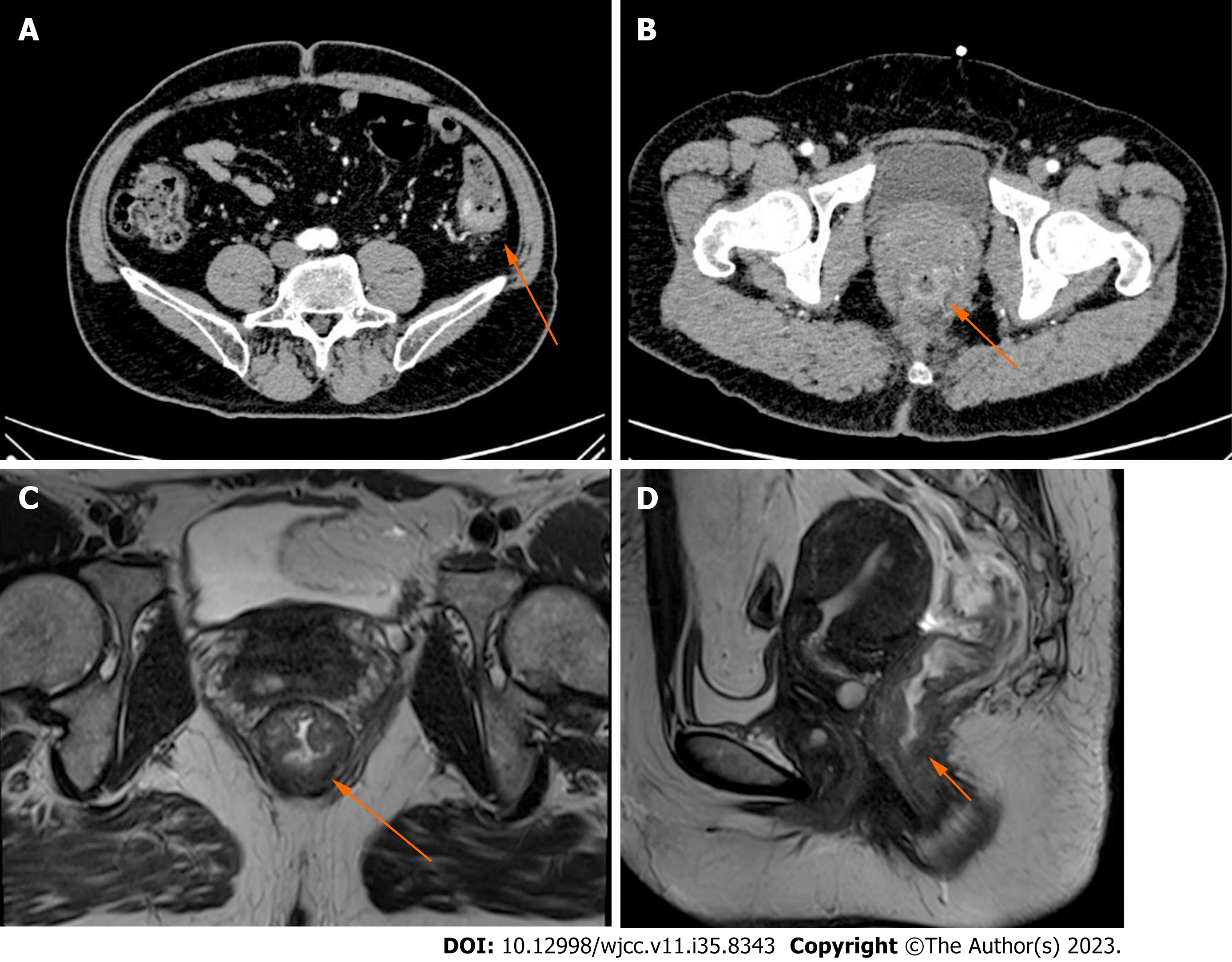
Figure 1 Computed tomography images.
A: Abdominal computed tomography (CT) revealed intestinal stenosis in the splenic flexure of the colon (arrow); B: Abdominal CT revealed obvious bowel wall thickness in the lower portions of the rectum (arrow); C and D: Axial and sagittal magnetic resonance imaging of the rectum depicted a sign of concentric tumor rings within the rectal wall and a tumor penetrating the layer of the intrinsic muscle into the perirectal mesenteric fat (arrow).
- Citation: Li F, Zhao B, Zhang L, Chen GQ, Zhu L, Feng XL, Yao H, Tang XF, Yang H, Liu YQ. Rare synchronous colorectal carcinoma with three pathological subtypes: A case report and review of the literature. World J Clin Cases 2023; 11(35): 8343-8349
- URL: https://www.wjgnet.com/2307-8960/full/v11/i35/8343.htm
- DOI: https://dx.doi.org/10.12998/wjcc.v11.i35.8343


