Copyright
©The Author(s) 2023.
World J Clin Cases. Nov 16, 2023; 11(32): 7926-7934
Published online Nov 16, 2023. doi: 10.12998/wjcc.v11.i32.7926
Published online Nov 16, 2023. doi: 10.12998/wjcc.v11.i32.7926
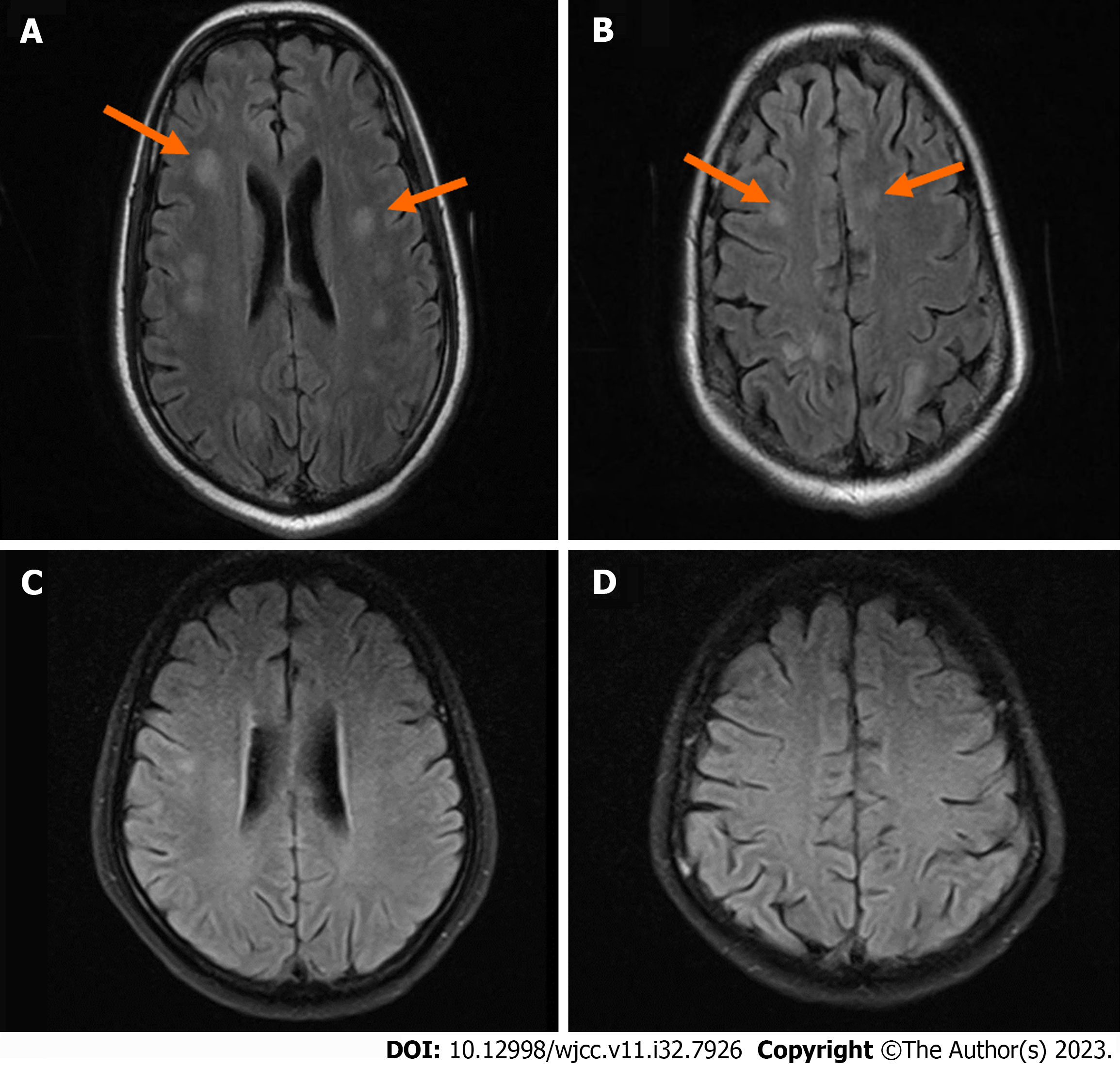
Figure 5 Brain magnetic resonance imaging.
A and B: Brain magnetic resonance imaging (MRI) of 2021-05-11 (orange arrows); C and D: Brain MRI of 2021-07-09 2021-05-11 brain MRI scan plus: T2 flair enhancement revealed multiple ischemic lesions in the right basal ganglia, bilateral frontal lobes, peri-ventricular, radiative crown, and hemioval center. 2021-07-09 brain MRI indicates improvement in the brain after treatment.
- Citation: Zheng JH, Wu D, Guo XY. Intracranial infection accompanied sweet’s syndrome in a patient with anti-interferon-γ autoantibodies: A case report. World J Clin Cases 2023; 11(32): 7926-7934
- URL: https://www.wjgnet.com/2307-8960/full/v11/i32/7926.htm
- DOI: https://dx.doi.org/10.12998/wjcc.v11.i32.7926


