Copyright
©The Author(s) 2023.
World J Clin Cases. Nov 16, 2023; 11(32): 7926-7934
Published online Nov 16, 2023. doi: 10.12998/wjcc.v11.i32.7926
Published online Nov 16, 2023. doi: 10.12998/wjcc.v11.i32.7926
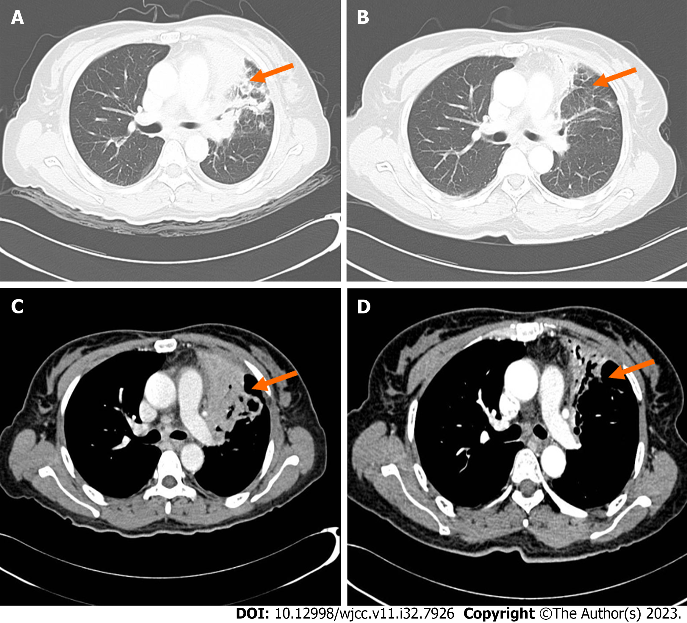
Figure 4 Chest computed tomography enhancement.
A: Lung window of 2021-05-10 (orange arrow); B: Lung window of 2021-07-12 (orange arrow); C: Mediastinal fenestra of 2021-05-10 (orange arrow); D: Mediastinal fenestra of 2021-07-12 2021-05-10 chest computed tomography (CT) revealed a high-density mass of size 7.7 cm × 7.2 cm × 5.9 cm in the upper lobe of the left lung in the lung window and lymph node enlargement in the mediastinal window. 2021-07-12 chest CT indicates improvement in the pulmonary lesion after treatment (orange arrow).
- Citation: Zheng JH, Wu D, Guo XY. Intracranial infection accompanied sweet’s syndrome in a patient with anti-interferon-γ autoantibodies: A case report. World J Clin Cases 2023; 11(32): 7926-7934
- URL: https://www.wjgnet.com/2307-8960/full/v11/i32/7926.htm
- DOI: https://dx.doi.org/10.12998/wjcc.v11.i32.7926


