Copyright
©The Author(s) 2023.
World J Clin Cases. Nov 16, 2023; 11(32): 7852-7857
Published online Nov 16, 2023. doi: 10.12998/wjcc.v11.i32.7852
Published online Nov 16, 2023. doi: 10.12998/wjcc.v11.i32.7852
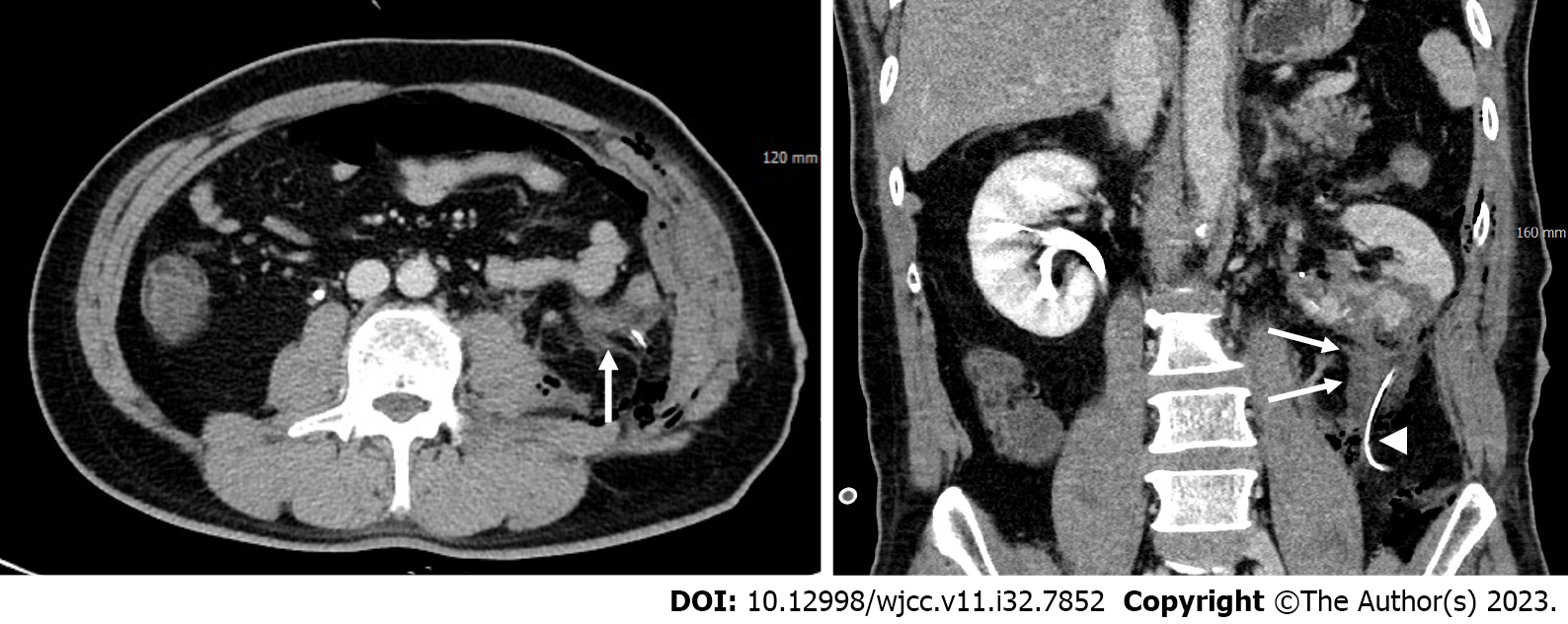
Figure 2 Post-operative computed tomography.
Post-operative computed tomography images obtained 1 d after partial nephrectomy revealing a small amount of fluid (arrows) in the inferior aspect of the left kidney, adjacent to the drainage tube (arrowhead), without evidence of contrast extravasation.
- Citation: Youm J, Choi MJ, Kim BM, Seo Y. Transcatheter embolization for hemorrhage from aberrant testicular artery after partial nephrectomy: A case report. World J Clin Cases 2023; 11(32): 7852-7857
- URL: https://www.wjgnet.com/2307-8960/full/v11/i32/7852.htm
- DOI: https://dx.doi.org/10.12998/wjcc.v11.i32.7852


