Copyright
©The Author(s) 2023.
World J Clin Cases. Nov 6, 2023; 11(31): 7718-7723
Published online Nov 6, 2023. doi: 10.12998/wjcc.v11.i31.7718
Published online Nov 6, 2023. doi: 10.12998/wjcc.v11.i31.7718
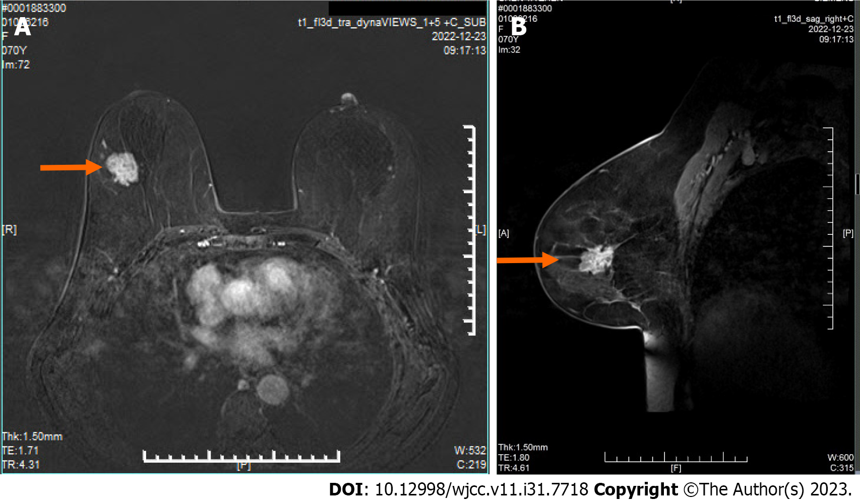
Figure 1 Dynamic contrast enhanced magnetic resonance imaging results.
A mass-like abnormal signal in the upper outer quadrant and outer upper quadrant. The size is approximately 29 mm × 21 mm × 25 mm, with irregular margins and spicules. A: Represents the sagittal view; B: Represents the coronal view.
- Citation: Li T, Zhang WH, Liu J, Mao YL, Liu S. Isolated axillary tumor deposit consistent with primary breast carcinoma: A case report. World J Clin Cases 2023; 11(31): 7718-7723
- URL: https://www.wjgnet.com/2307-8960/full/v11/i31/7718.htm
- DOI: https://dx.doi.org/10.12998/wjcc.v11.i31.7718


