Copyright
©The Author(s) 2023.
World J Clin Cases. Nov 6, 2023; 11(31): 7663-7672
Published online Nov 6, 2023. doi: 10.12998/wjcc.v11.i31.7663
Published online Nov 6, 2023. doi: 10.12998/wjcc.v11.i31.7663
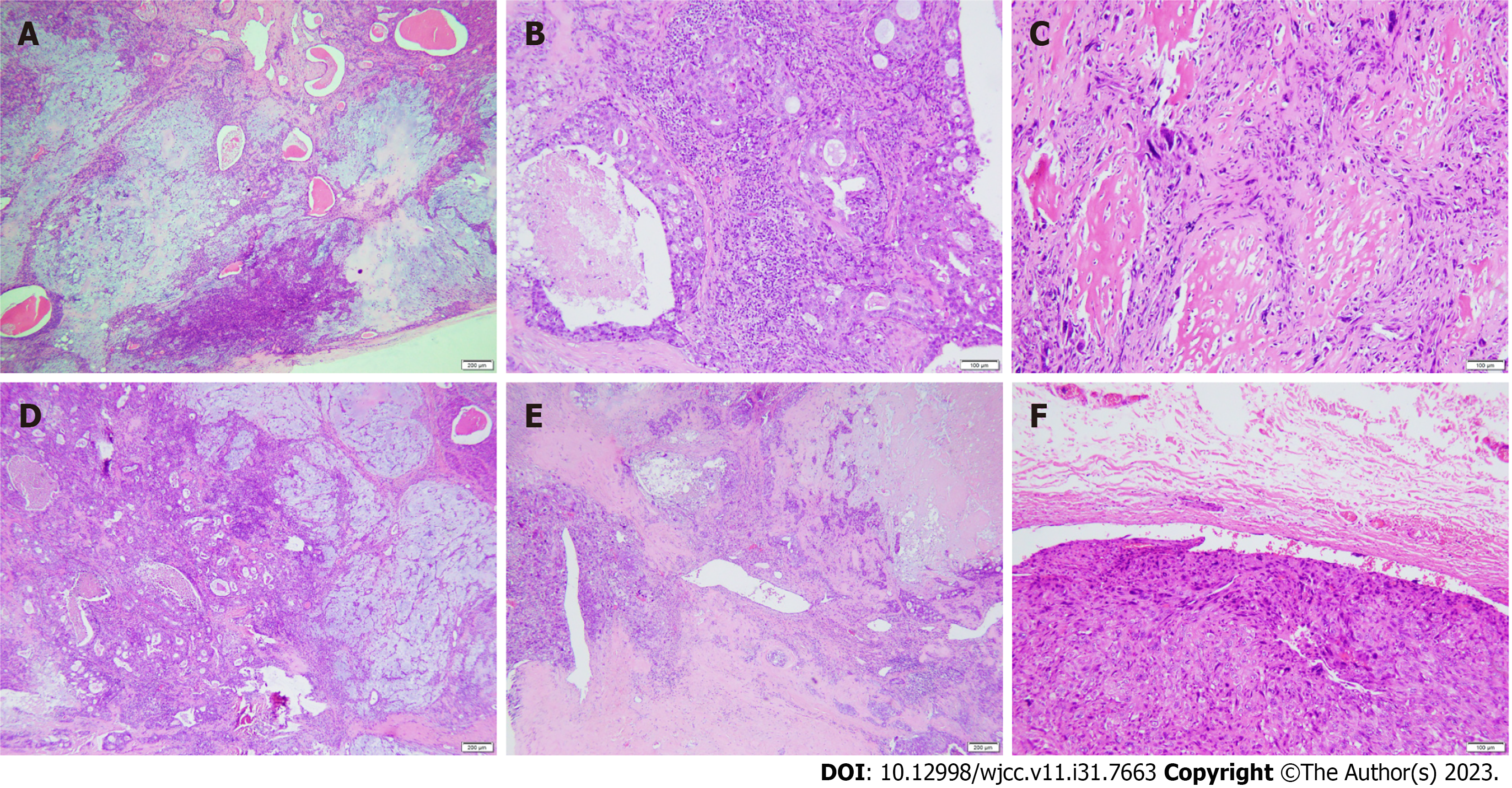
Figure 3 Histological features of carcinosarcoma (hematoxylin and eosin staining).
A: The pleomorphic adenoma region had a rich tissue structure and mild cell morphological changes; B: In the salivary duct carcinoma area, comedo-like necrosis was seen in the center of the solid epithelial mass; C: In the sarcomatous area, the cells showed obvious atypia and abundant bone matrix formation around the cells; D: Pleomorphic adenoma (right) mixed with salivary duct carcinoma (left); E: Salivary duct carcinoma (right) mixed with osteosarcoma (left); F: A tumor thrombus was found in the venous lumen.
- Citation: Tang YY, Zhu GQ, Zheng ZJ, Yao LH, Wan ZX, Liang XH, Tang YL. Carcinosarcoma of the deep lobe of the parotid gland in the parapharyngeal region: A case report. World J Clin Cases 2023; 11(31): 7663-7672
- URL: https://www.wjgnet.com/2307-8960/full/v11/i31/7663.htm
- DOI: https://dx.doi.org/10.12998/wjcc.v11.i31.7663


