Copyright
©The Author(s) 2023.
World J Clin Cases. Nov 6, 2023; 11(31): 7663-7672
Published online Nov 6, 2023. doi: 10.12998/wjcc.v11.i31.7663
Published online Nov 6, 2023. doi: 10.12998/wjcc.v11.i31.7663
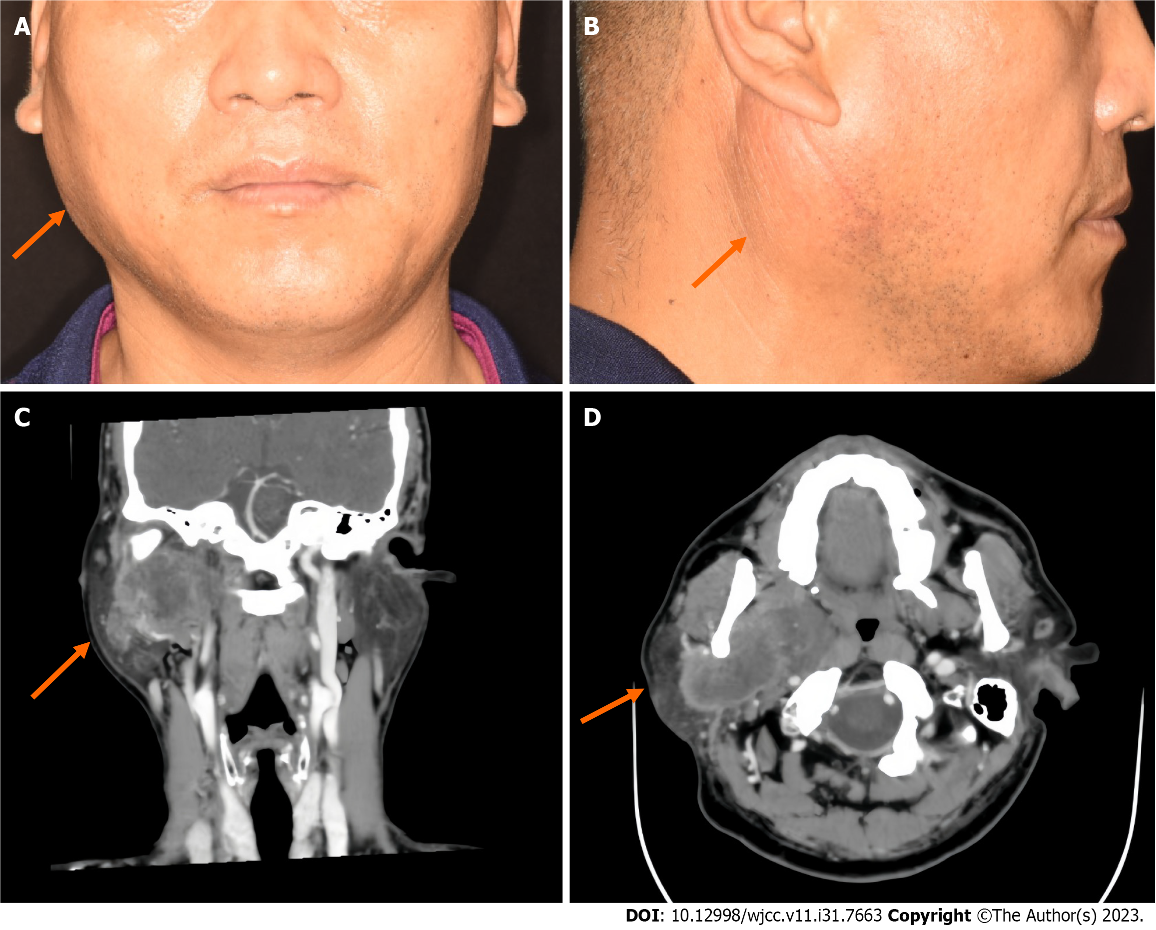
Figure 1 Preoperative photographs and enhanced computed tomography images of the patient.
A: Front; B: Side; C: Computed tomography of the right parotid gland revealed a mass in its deep lobe that also extended into the parapharynx; D: There was obvious uneven enhancement, with local ring enhancement.
- Citation: Tang YY, Zhu GQ, Zheng ZJ, Yao LH, Wan ZX, Liang XH, Tang YL. Carcinosarcoma of the deep lobe of the parotid gland in the parapharyngeal region: A case report. World J Clin Cases 2023; 11(31): 7663-7672
- URL: https://www.wjgnet.com/2307-8960/full/v11/i31/7663.htm
- DOI: https://dx.doi.org/10.12998/wjcc.v11.i31.7663


