Copyright
©The Author(s) 2023.
World J Clin Cases. Nov 6, 2023; 11(31): 7629-7634
Published online Nov 6, 2023. doi: 10.12998/wjcc.v11.i31.7629
Published online Nov 6, 2023. doi: 10.12998/wjcc.v11.i31.7629
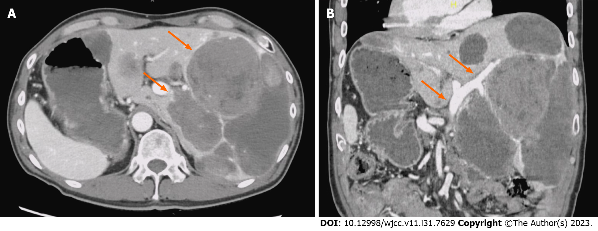
Figure 1 Computed tomography of the abdomen and pelvis with contrast images.
A: Computed tomography (CT) image of the abdomen and pelvis with contrast. Axial section shows situs inversus and increased liver metastases (orange arrows); B: CT image of the abdomen and pelvis with contrast. Coronal section shows no portal vein obstruction (orange arrows) or significant portosystemic shunt.
- Citation: Hayakawa T, Funakoshi S, Hamamoto Y, Hirata K, Kanai T. Sunitinib-induced hyperammonemic encephalopathy in metastatic gastrointestinal stromal tumors: A case report. World J Clin Cases 2023; 11(31): 7629-7634
- URL: https://www.wjgnet.com/2307-8960/full/v11/i31/7629.htm
- DOI: https://dx.doi.org/10.12998/wjcc.v11.i31.7629


