Copyright
©The Author(s) 2023.
World J Clin Cases. Nov 6, 2023; 11(31): 7570-7582
Published online Nov 6, 2023. doi: 10.12998/wjcc.v11.i31.7570
Published online Nov 6, 2023. doi: 10.12998/wjcc.v11.i31.7570
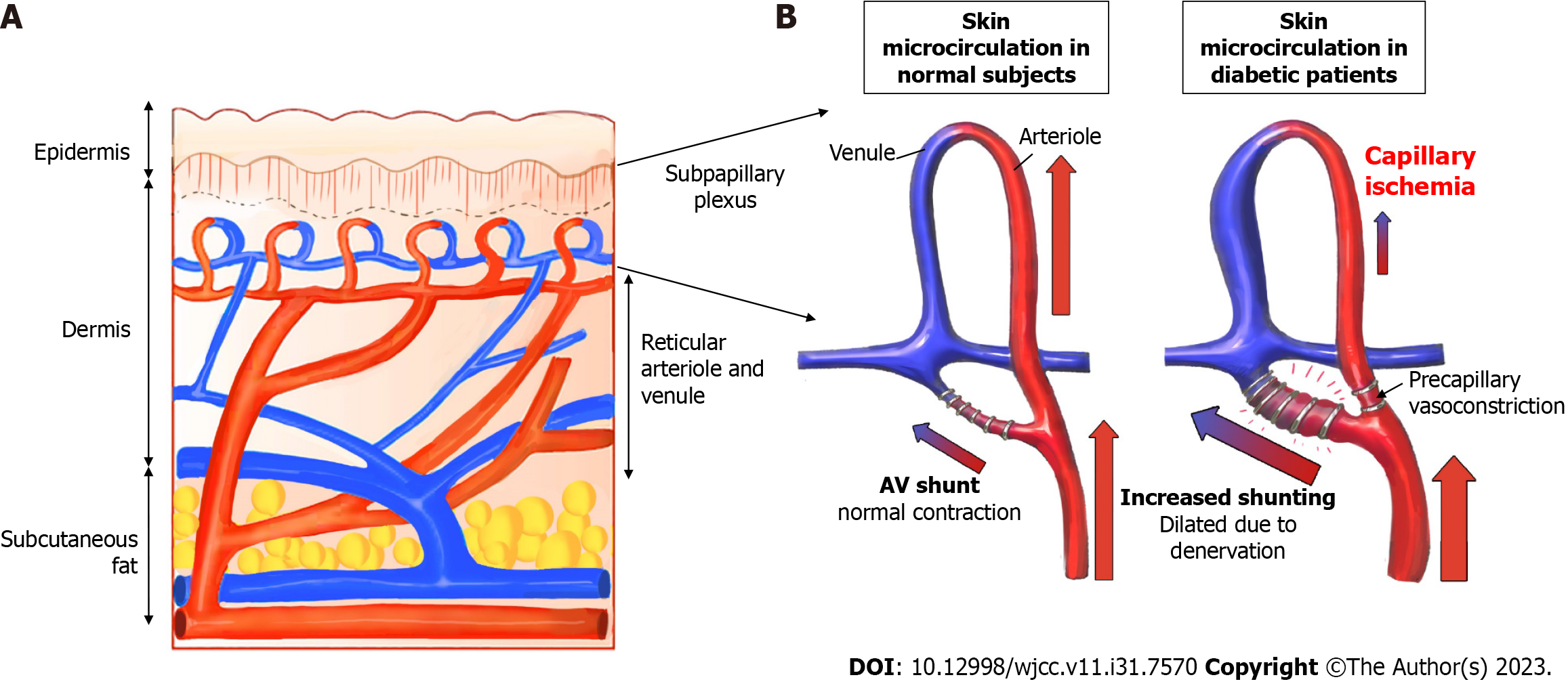
Figure 8 Schematic illustration of the increase in a subpapillary arteriovenous shunt in diabetic patients.
A: Cross-sectional structure of the skin; the subpapillary plexus is located in the lower layer of the epidermis; B: In a patent with diabetes, arteriovenous shunt is increased, and periphery blood flow is reduced.
- Citation: Lee DW, Hwang YS, Byeon JY, Kim JH, Choi HJ. Does the advantage of transcutaneous oximetry measurements in diabetic foot ulcer apply equally to free flap reconstruction? World J Clin Cases 2023; 11(31): 7570-7582
- URL: https://www.wjgnet.com/2307-8960/full/v11/i31/7570.htm
- DOI: https://dx.doi.org/10.12998/wjcc.v11.i31.7570


