Copyright
©The Author(s) 2023.
World J Clin Cases. Oct 26, 2023; 11(30): 7485-7491
Published online Oct 26, 2023. doi: 10.12998/wjcc.v11.i30.7485
Published online Oct 26, 2023. doi: 10.12998/wjcc.v11.i30.7485
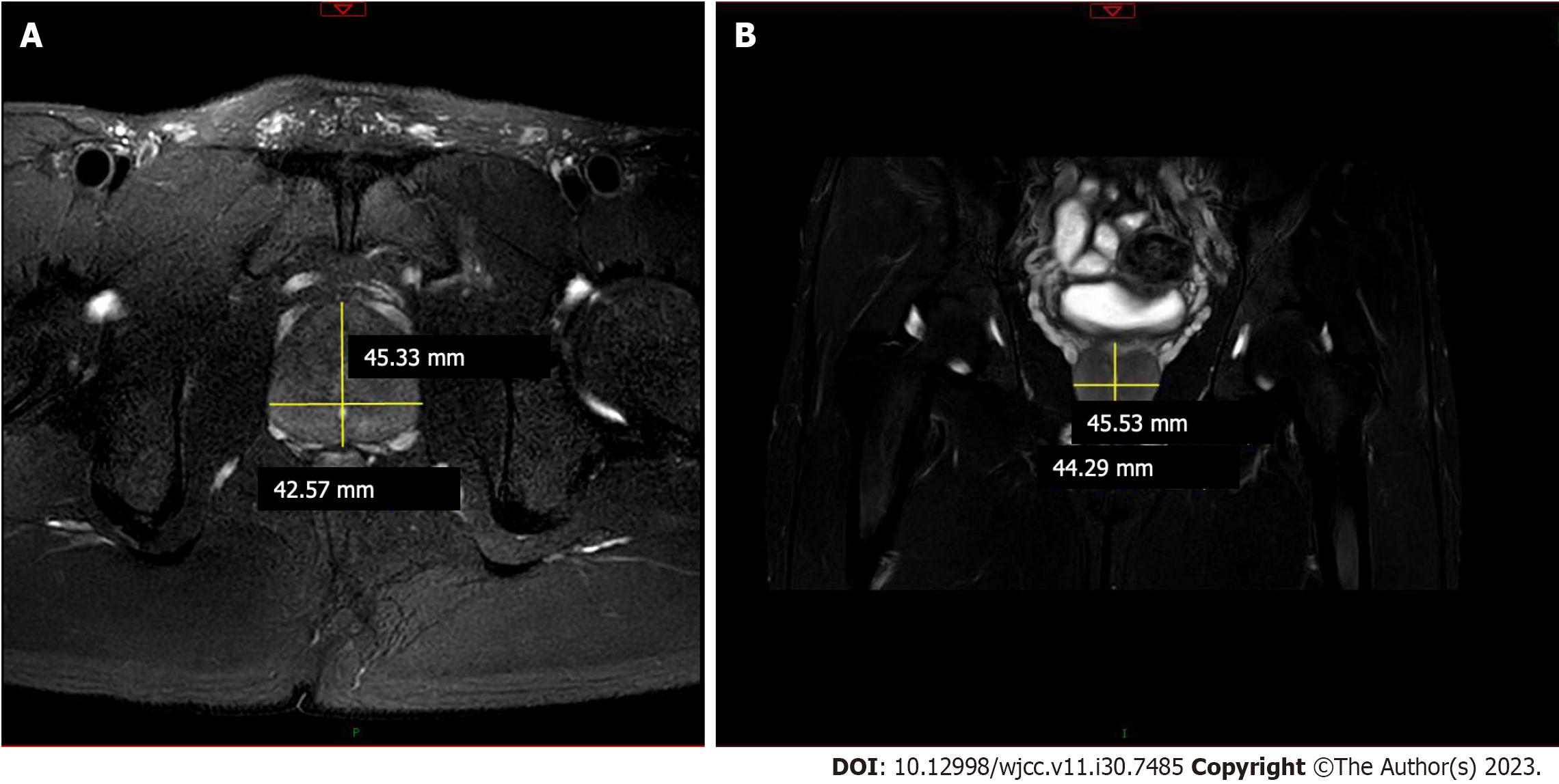
Figure 4 Magnetic resonance imaging of the prostate gland.
A: Prostate: Prostate enlargement, about 46 mm × 41 mm. The boundary between the central gland and the peripheral zone of the prostate was unclear. T2WI signal decreased diffusely; B: Enhanced scanning of the prostate showed uneven enhancement. The fat gap around the prostate was clear.
- Citation: Yu Y, Wang QQ, Jian L, Yang DC. Infrequent organ involvement in immunoglobulin G4-related prostate disease: A case report. World J Clin Cases 2023; 11(30): 7485-7491
- URL: https://www.wjgnet.com/2307-8960/full/v11/i30/7485.htm
- DOI: https://dx.doi.org/10.12998/wjcc.v11.i30.7485


