Copyright
©The Author(s) 2023.
World J Clin Cases. Oct 26, 2023; 11(30): 7485-7491
Published online Oct 26, 2023. doi: 10.12998/wjcc.v11.i30.7485
Published online Oct 26, 2023. doi: 10.12998/wjcc.v11.i30.7485
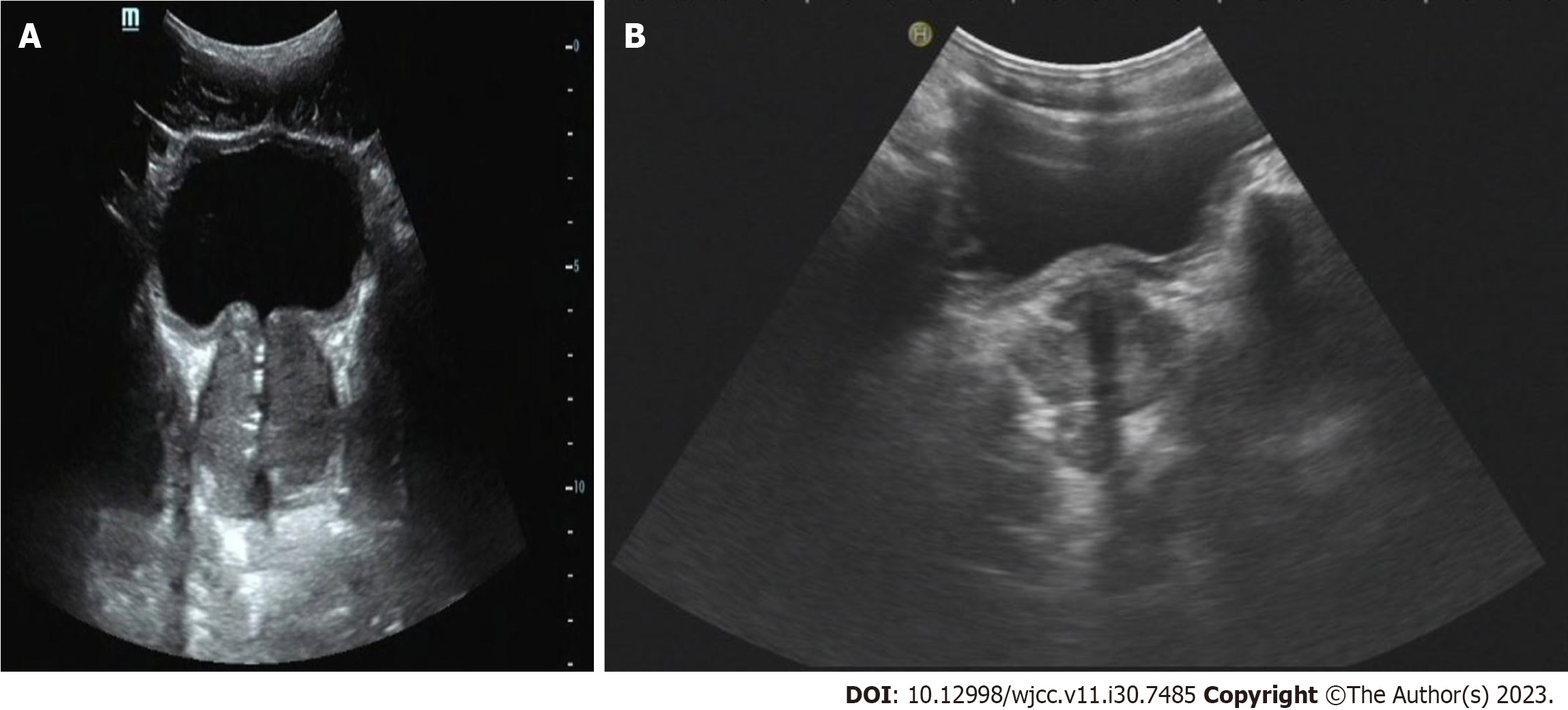
Figure 3 Ultrasound of the prostate.
A: The prostate section was 5.5 cm × 4.1 cm × 4.6 cm in size, full in shape, and showed uneven distribution of internal light spots. A few strong echo spots were observed; B: The prostate section was 3.5 cm × 3.2 cm × 3.0 cm in size 14 mo later.
- Citation: Yu Y, Wang QQ, Jian L, Yang DC. Infrequent organ involvement in immunoglobulin G4-related prostate disease: A case report. World J Clin Cases 2023; 11(30): 7485-7491
- URL: https://www.wjgnet.com/2307-8960/full/v11/i30/7485.htm
- DOI: https://dx.doi.org/10.12998/wjcc.v11.i30.7485


