Copyright
©The Author(s) 2023.
World J Clin Cases. Oct 26, 2023; 11(30): 7403-7412
Published online Oct 26, 2023. doi: 10.12998/wjcc.v11.i30.7403
Published online Oct 26, 2023. doi: 10.12998/wjcc.v11.i30.7403
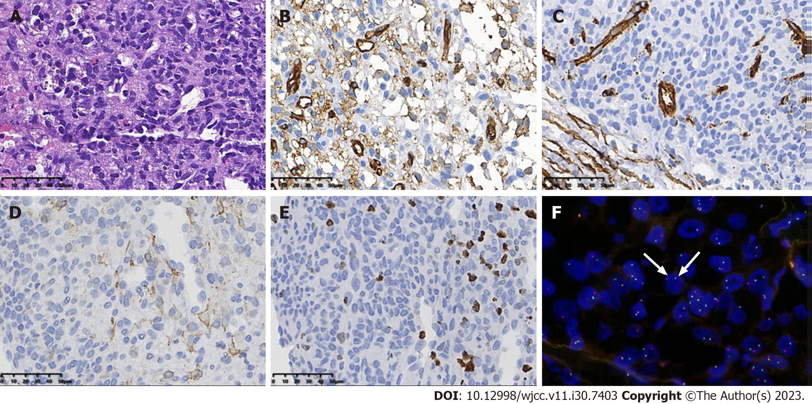
Figure 6 Pathological findings of the congenital infantile fibrosarcoma in Case 1.
A: H&E staining image. The tumor cells showing eosinophilic, diffuse sheets of monotonous round cells with fine chromatin, and indistinct nucleoli; B-D: Immunochemistry demonstrated the expression of smooth muscle actin, CD34, and CD31 in the tumor cells; E: The immunochemical study showed that the Ki-67 proliferation index was approximately 20%; F: Using fluorescence in situ hybridization, the tumor cells showed NTRK3 gene rearrangement, with break-apart green (telomeric) and red (centromeric) signals (white arrows).
- Citation: Liang RN, Jiang J, Zhang J, Liu X, Ma MY, Liu QL, Ma L, Zhou L, Wang Y, Wang J, Zhou Q, Yu SS. Prenatal ultrasound diagnosis of congenital infantile fibrosarcoma and congenital hemangioma: Three case reports. World J Clin Cases 2023; 11(30): 7403-7412
- URL: https://www.wjgnet.com/2307-8960/full/v11/i30/7403.htm
- DOI: https://dx.doi.org/10.12998/wjcc.v11.i30.7403


