Copyright
©The Author(s) 2023.
World J Clin Cases. Oct 26, 2023; 11(30): 7403-7412
Published online Oct 26, 2023. doi: 10.12998/wjcc.v11.i30.7403
Published online Oct 26, 2023. doi: 10.12998/wjcc.v11.i30.7403
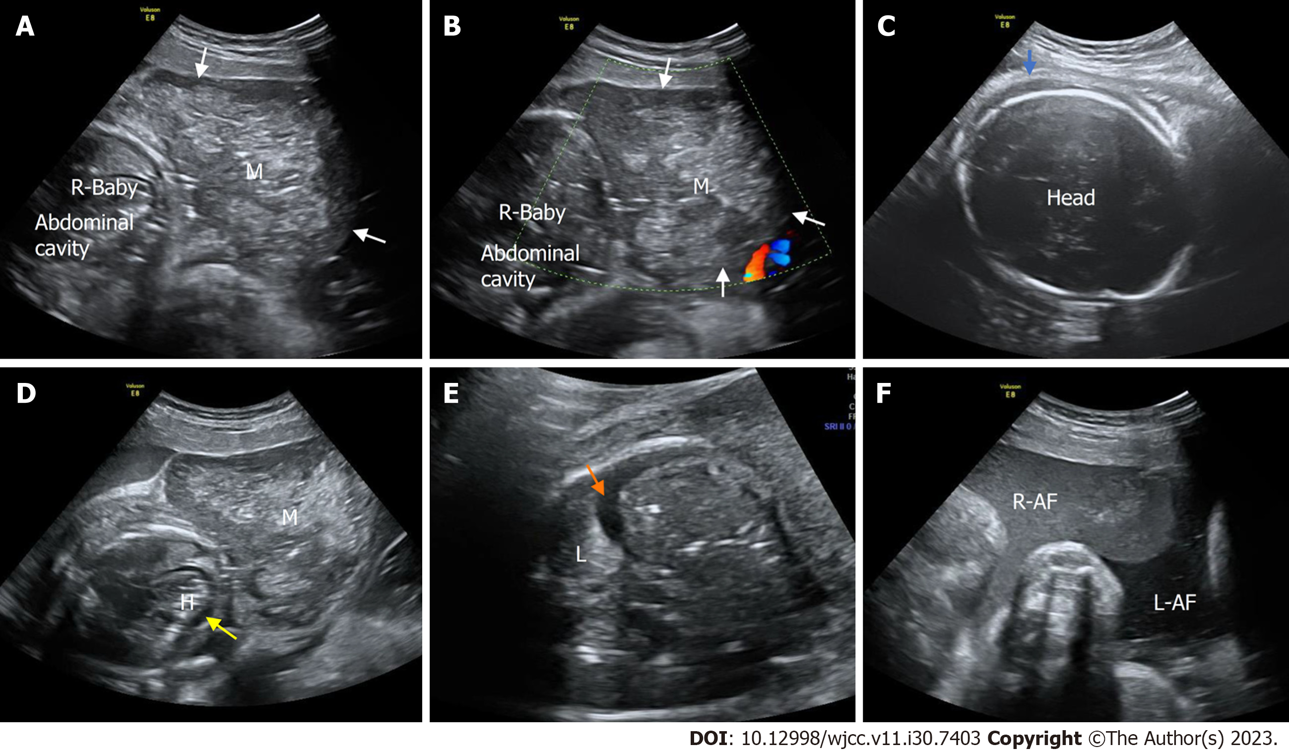
Figure 5 Prenatal ultrasonic imaging of the congenital hemangioma of the affected twin fetus in Case 3 at 33+0 wk of gestation.
A: Two-dimensional ultrasound of the abdominal horizontal cross-section of the mass of the affected fetus revealing that the mass was enlarged and heterogeneous; B: CDFI of an abdominal horizontal cross-section of the mass showing no blood flow signal within the mass. M: Mass; R: Right; white arrow, the mass; C: A two-dimensional ultrasound image of the fetal head showing subcutaneous edema (blue arrow); D: A two-dimensional transverse section of the fetal chest showing intrapericardial fluid collection (yellow arrow). H, heart; M, mass; E: A two-dimensional thoracic horizontal cross-section of the affected fetus showing fetal pleural effusion (orange arrow). L: Lung; M: Mass; R: Right; white arrow, the mass; F: Two amniotic cavities on two-dimensional ultrasound showing that the amniotic fluid of the affected fetus (R-AF) was not clear. R-AF: Right amniotic fluid. LAF left amniotic fluid.
- Citation: Liang RN, Jiang J, Zhang J, Liu X, Ma MY, Liu QL, Ma L, Zhou L, Wang Y, Wang J, Zhou Q, Yu SS. Prenatal ultrasound diagnosis of congenital infantile fibrosarcoma and congenital hemangioma: Three case reports. World J Clin Cases 2023; 11(30): 7403-7412
- URL: https://www.wjgnet.com/2307-8960/full/v11/i30/7403.htm
- DOI: https://dx.doi.org/10.12998/wjcc.v11.i30.7403


