Copyright
©The Author(s) 2023.
World J Clin Cases. Oct 26, 2023; 11(30): 7403-7412
Published online Oct 26, 2023. doi: 10.12998/wjcc.v11.i30.7403
Published online Oct 26, 2023. doi: 10.12998/wjcc.v11.i30.7403
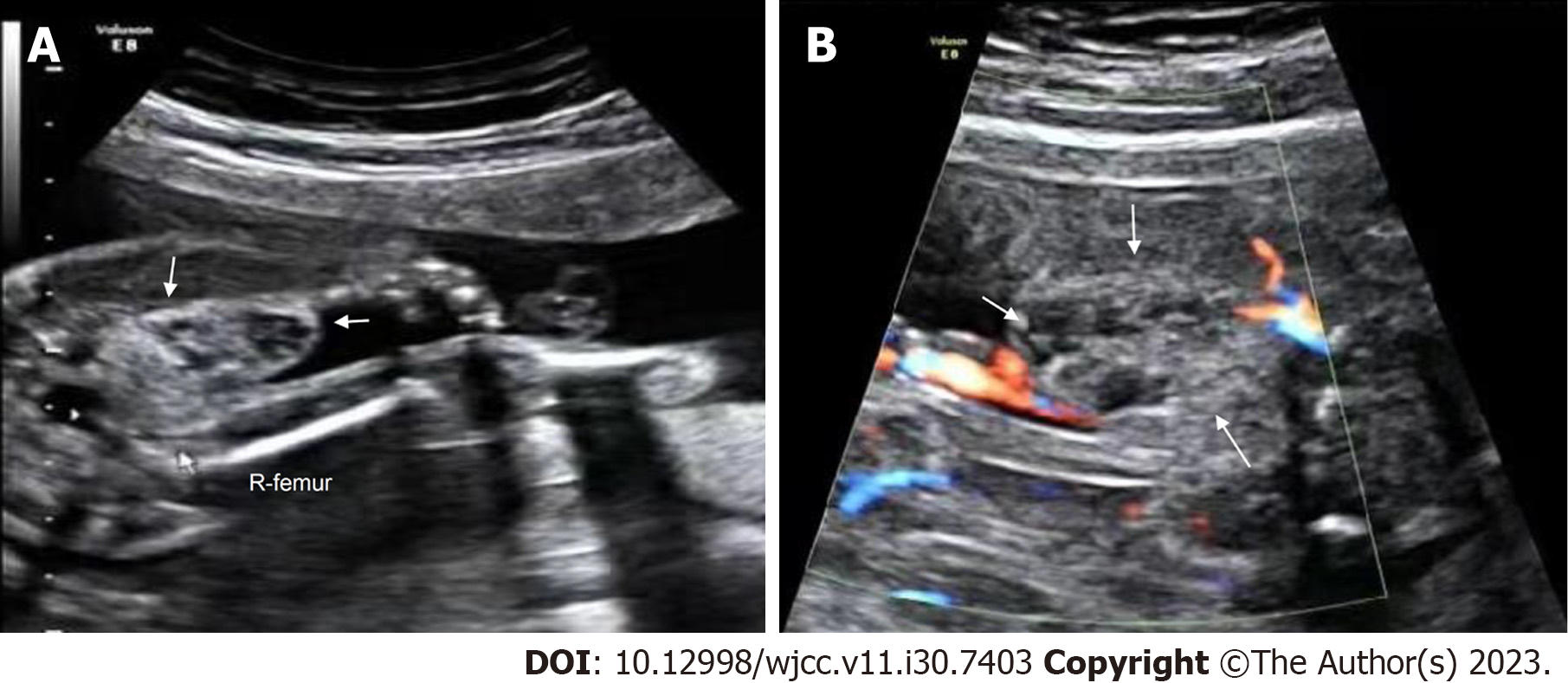
Figure 3 Prenatal ultrasonic imaging of the congenital hemangioma in Case 2.
A: Prenatal two-dimensional ultrasound image revealing a well-defined, heterogeneous mass at the root of the right thigh of the fetus at 23+1 wk of gestation. White arrow, the mass; R-femur, the right femur of the fetus; B: CDFI of the mass at the root of the right thigh of the fetus at 23+1 wk of gestation showing sparse punctate blood flow around the mass. White arrow, the mass.
- Citation: Liang RN, Jiang J, Zhang J, Liu X, Ma MY, Liu QL, Ma L, Zhou L, Wang Y, Wang J, Zhou Q, Yu SS. Prenatal ultrasound diagnosis of congenital infantile fibrosarcoma and congenital hemangioma: Three case reports. World J Clin Cases 2023; 11(30): 7403-7412
- URL: https://www.wjgnet.com/2307-8960/full/v11/i30/7403.htm
- DOI: https://dx.doi.org/10.12998/wjcc.v11.i30.7403


