Copyright
©The Author(s) 2023.
World J Clin Cases. Oct 26, 2023; 11(30): 7403-7412
Published online Oct 26, 2023. doi: 10.12998/wjcc.v11.i30.7403
Published online Oct 26, 2023. doi: 10.12998/wjcc.v11.i30.7403
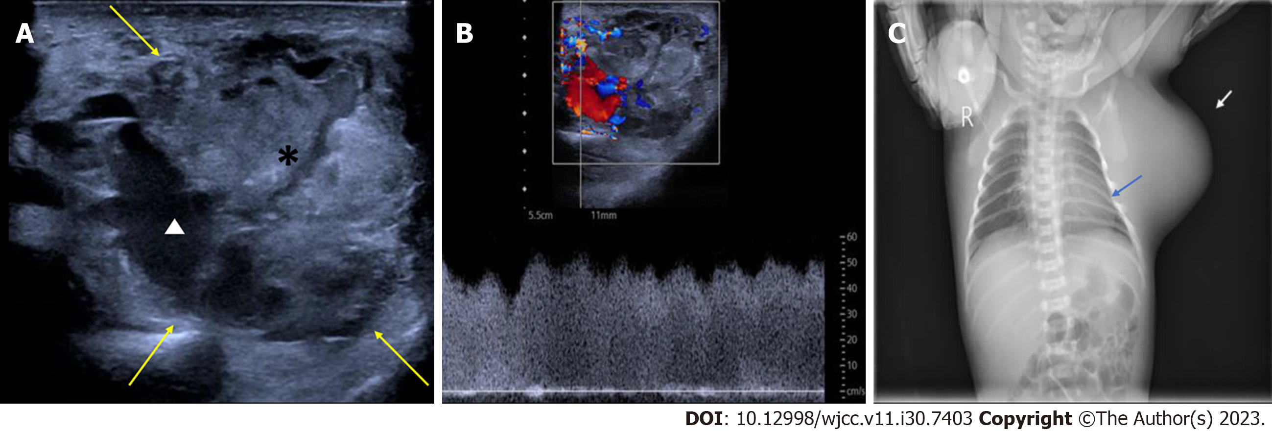
Figure 2 Postnatal imaging of the congenital infantile fibrosarcoma in Case 1.
A: One-day postnatal two-dimensional ultrasound using a linear array transducer with a frequency of 5-12 MHz, revealing a well-defined mass in the left axilla consisting mainly of isoechoic parenchymal components (*) with a few anechoic areas (white triangle) within the mass. Yellow arrow, the boundary of the mass; B: One-day postnatal pulse Doppler ultrasound revealing a rich blood flow and low resistance blood flow spectrum in the mass; C: Postnatal X-ray of the newborn showed a soft tissue density shadow protruding from the left axilla and lateral chest wall, resulting in compression of the adjacent ribs (blue arrow). White arrow, the mass on X-ray.
- Citation: Liang RN, Jiang J, Zhang J, Liu X, Ma MY, Liu QL, Ma L, Zhou L, Wang Y, Wang J, Zhou Q, Yu SS. Prenatal ultrasound diagnosis of congenital infantile fibrosarcoma and congenital hemangioma: Three case reports. World J Clin Cases 2023; 11(30): 7403-7412
- URL: https://www.wjgnet.com/2307-8960/full/v11/i30/7403.htm
- DOI: https://dx.doi.org/10.12998/wjcc.v11.i30.7403


