Copyright
©The Author(s) 2023.
World J Clin Cases. Jan 26, 2023; 11(3): 700-708
Published online Jan 26, 2023. doi: 10.12998/wjcc.v11.i3.700
Published online Jan 26, 2023. doi: 10.12998/wjcc.v11.i3.700
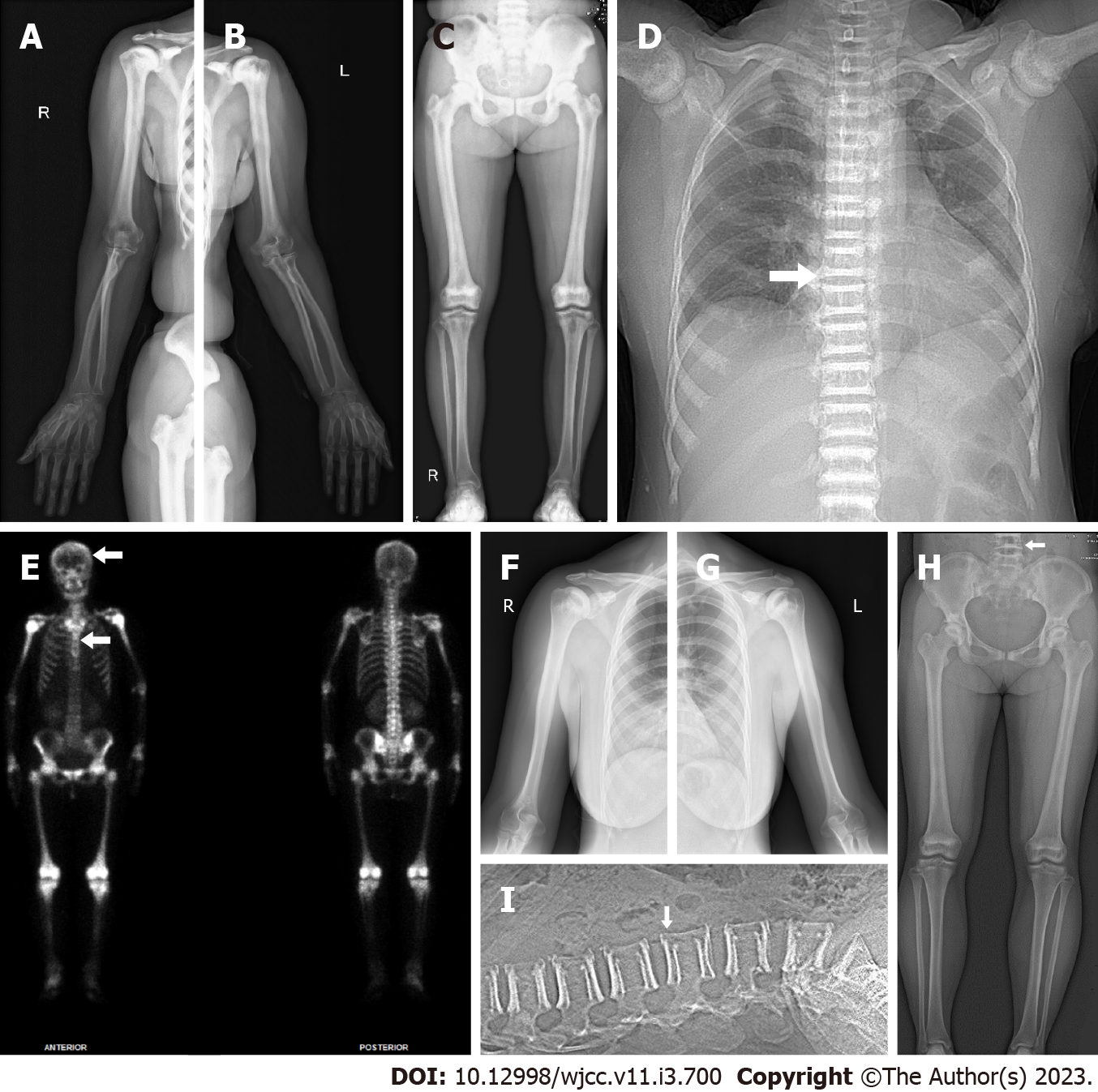
Figure 2 The imaging examination of the proband and the proband’s daughter.
A-C: X-ray image of the proband (II:4) demonstrated increased bone density in the right upper extremity (A), the left upper extremity (B) and the lower extremities (C); D: The chest X-ray of the proband (II:4) showed increased bone density of pelvic, with a “bone-within-bone” appearance in lateral of the spine, indicating classic vertebral endplate thickening (white arrow indicates the “sandwich vertebrae” sign); E: Whole-body bone technetium-99m single-photon emission computed tomography scan revealed extremely high bone uptake of long bone, ribs, and spine with absent renal radioactivity visualization (white arrows indicate “helmet” and “tie” sign); F and G: X-ray image of the proband’s daughter (III:1) showed that the bone density of the bone tip, the humerus and the ribs and the medullary cavity became narrow in the right upper extremity (F) and the left upper extremity(G); H and I: X-ray image of the proband’s daughter (III:1) in the lower extremities (H) and the lumbar vertebra (I) indicated classic vertebral endplate thickening (white arrow indicates the “sandwich vertebrae” sign).
- Citation: Gong HP, Ren Y, Zha PP, Zhang WY, Zhang J, Zhang ZW, Wang C. Clinical and genetic diagnosis of autosomal dominant osteopetrosis type II in a Chinese family: A case report. World J Clin Cases 2023; 11(3): 700-708
- URL: https://www.wjgnet.com/2307-8960/full/v11/i3/700.htm
- DOI: https://dx.doi.org/10.12998/wjcc.v11.i3.700


