Copyright
©The Author(s) 2023.
World J Clin Cases. Jan 26, 2023; 11(3): 692-699
Published online Jan 26, 2023. doi: 10.12998/wjcc.v11.i3.692
Published online Jan 26, 2023. doi: 10.12998/wjcc.v11.i3.692
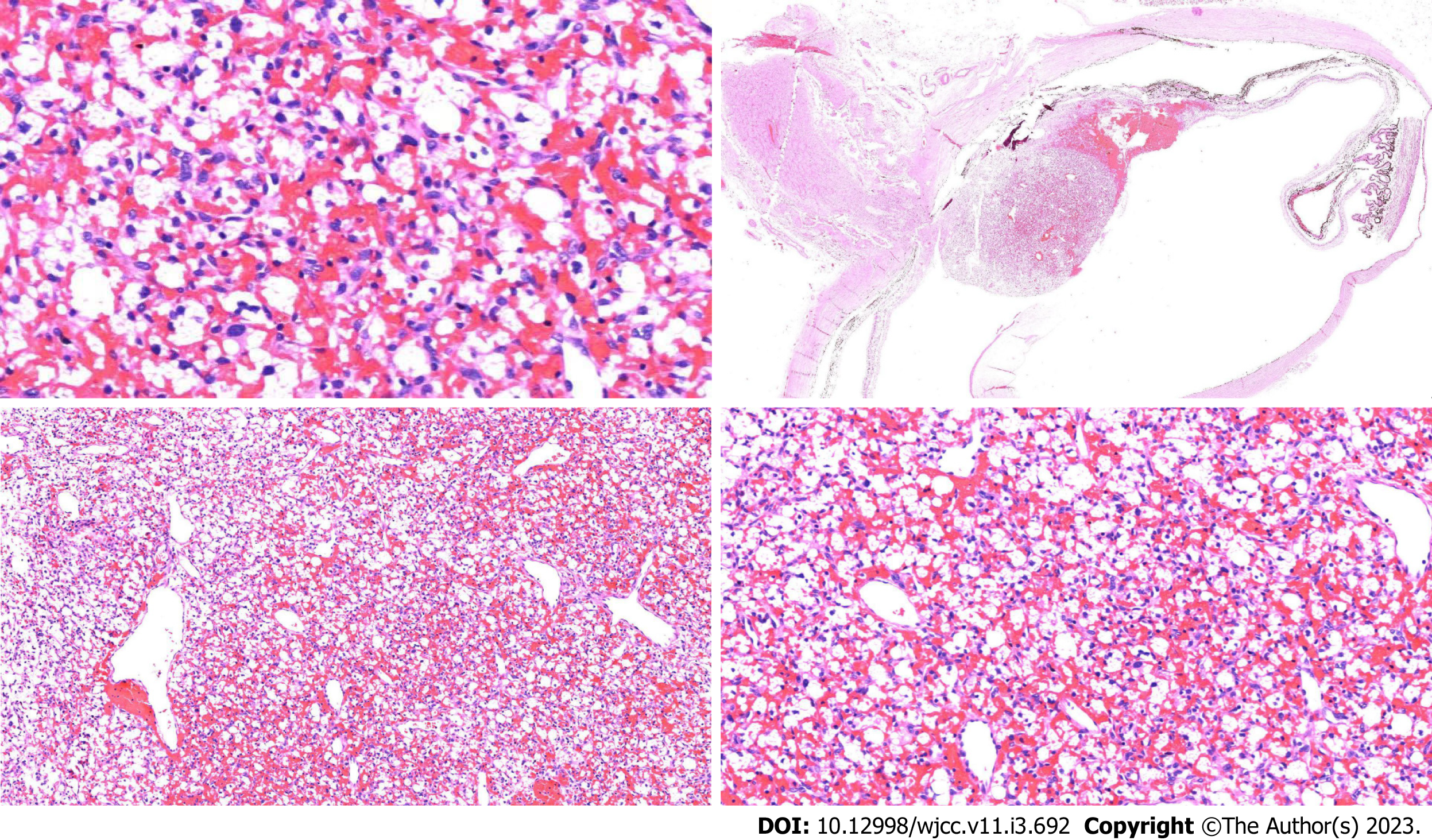
Figure 4 Postoperative histopathological and immunohistological images of left retinal hemangioblastoma.
The left eyeball lesions were mainly composed of two components, capillaries and interstitial cells surrounded by vacuolated or eosinophilic cytoplasm, which showed epithelioid stromal cells and staghorn dilated thin-walled vessels in capillaries (hematoxylin-eosin staining, magnification × 4).
- Citation: Tang X, Ji HM, Li WW, Ding ZX, Ye SL. Imaging features of retinal hemangioblastoma: A case report. World J Clin Cases 2023; 11(3): 692-699
- URL: https://www.wjgnet.com/2307-8960/full/v11/i3/692.htm
- DOI: https://dx.doi.org/10.12998/wjcc.v11.i3.692


