Copyright
©The Author(s) 2023.
World J Clin Cases. Jan 26, 2023; 11(3): 692-699
Published online Jan 26, 2023. doi: 10.12998/wjcc.v11.i3.692
Published online Jan 26, 2023. doi: 10.12998/wjcc.v11.i3.692
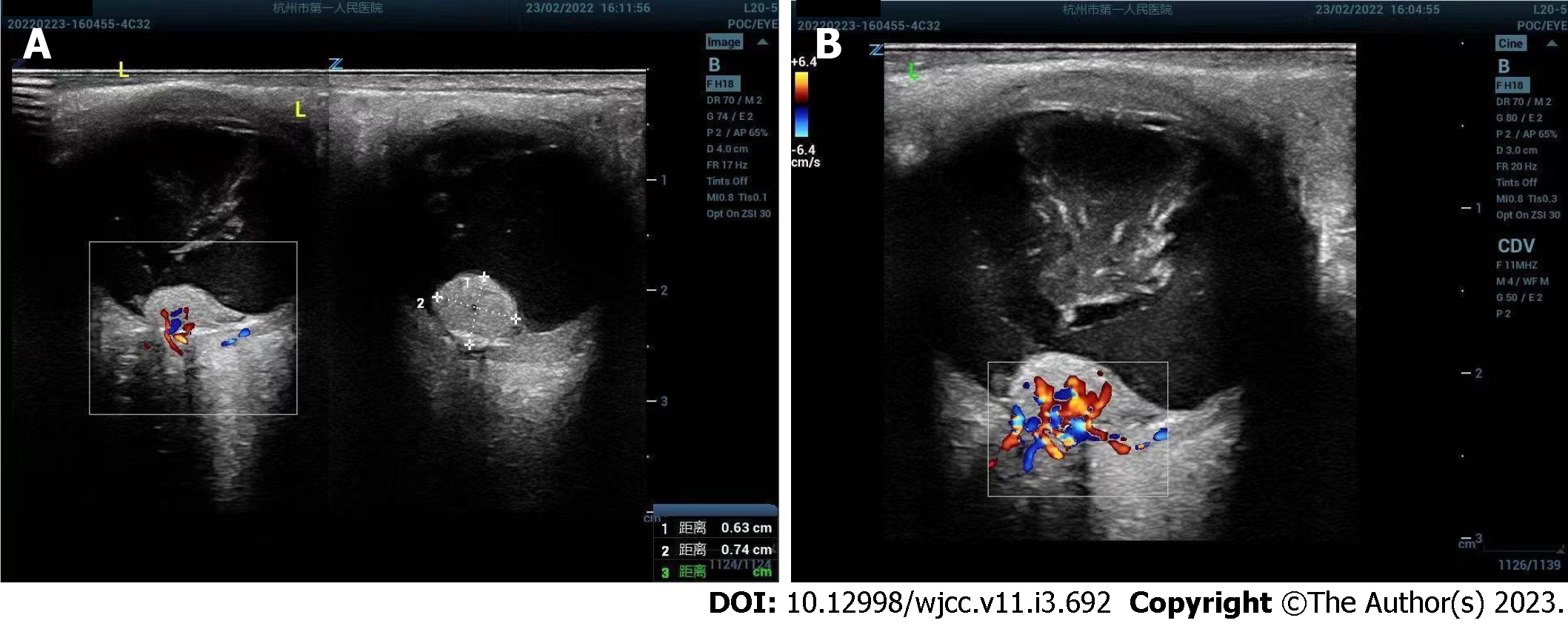
Figure 1 Ultrasound images of left retinal hemangioblastoma.
A: Ultrasound showed an irregular isoechoic mass of about 6.3 mm × 7.4 mm in front of the left optic nerve head; B: Color doppler flow imaging showed abundant blood flow signals in the lesion.
- Citation: Tang X, Ji HM, Li WW, Ding ZX, Ye SL. Imaging features of retinal hemangioblastoma: A case report. World J Clin Cases 2023; 11(3): 692-699
- URL: https://www.wjgnet.com/2307-8960/full/v11/i3/692.htm
- DOI: https://dx.doi.org/10.12998/wjcc.v11.i3.692


