Copyright
©The Author(s) 2023.
World J Clin Cases. Jan 26, 2023; 11(3): 684-691
Published online Jan 26, 2023. doi: 10.12998/wjcc.v11.i3.684
Published online Jan 26, 2023. doi: 10.12998/wjcc.v11.i3.684
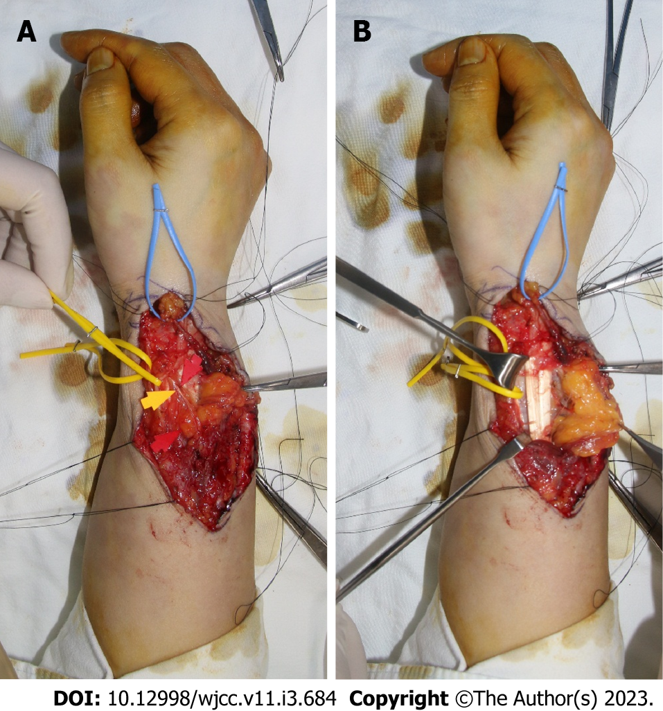
Figure 4 Intraoperative photographs.
A: The previous surgical scar was used, and an incision was applied along the run of the extensor pollicis brevis (EPB) to expose the lipomatous mass. A radial artery marked with yellow vessel loop and lipoma in the EPB tendon sheath marked with blue vessel loop were identified; B: Lipoma infiltrating into the muscle portion of EPB was removed while preserving the superficial radial nerve marked with yellow and red arrows.
- Citation: Byeon JY, Hwang YS, Lee JH, Choi HJ. Recurrent intramuscular lipoma at extensor pollicis brevis: A case report. World J Clin Cases 2023; 11(3): 684-691
- URL: https://www.wjgnet.com/2307-8960/full/v11/i3/684.htm
- DOI: https://dx.doi.org/10.12998/wjcc.v11.i3.684


