Copyright
©The Author(s) 2023.
World J Clin Cases. Jan 26, 2023; 11(3): 684-691
Published online Jan 26, 2023. doi: 10.12998/wjcc.v11.i3.684
Published online Jan 26, 2023. doi: 10.12998/wjcc.v11.i3.684
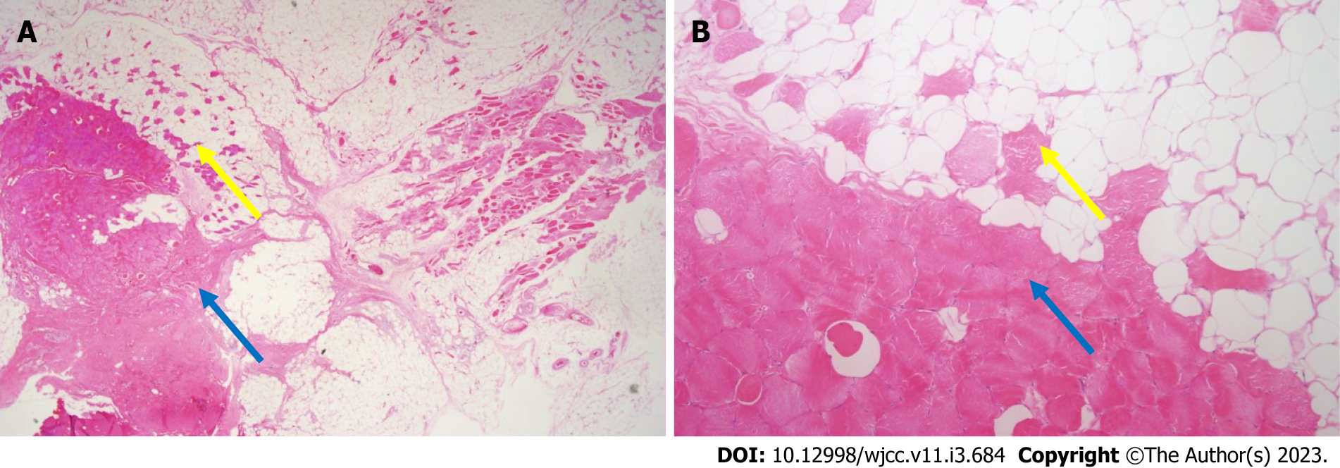
Figure 3 Histological findings.
A: Microscopic findings of intramuscular lipomas showing mature adipocytes and skeletal muscle fibers (pink color) (× 10.25 magnification, hematoxylin and eosin staining); B: Skeletal muscle fibers (pink color) are observed between mature adipocytes (× 100 magnification, hematoxylin and eosin staining). Yellow arrows indicate muscle components in fat. Blue arrows indicate muscle belly of extensor pollicis brevis.
- Citation: Byeon JY, Hwang YS, Lee JH, Choi HJ. Recurrent intramuscular lipoma at extensor pollicis brevis: A case report. World J Clin Cases 2023; 11(3): 684-691
- URL: https://www.wjgnet.com/2307-8960/full/v11/i3/684.htm
- DOI: https://dx.doi.org/10.12998/wjcc.v11.i3.684


