Copyright
©The Author(s) 2023.
World J Clin Cases. Jan 26, 2023; 11(3): 684-691
Published online Jan 26, 2023. doi: 10.12998/wjcc.v11.i3.684
Published online Jan 26, 2023. doi: 10.12998/wjcc.v11.i3.684
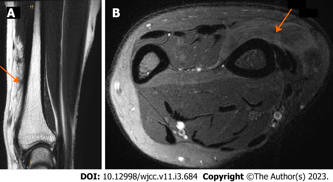
Figure 2 Preoperative magnetic resonance imaging showing lipomatous masses with fat-like attenuation in extensor pollicis brevis muscles and ligaments.
A: In the coronal view, lipomatous mass infiltrated in the muscle was identified. Boundaries with surrounding muscles and ligaments were unclear; B: In the axial view, lipomatous mass of similar attenuate to fat was identified. An orange arrow indicates a lipomatous mass that infiltrates muscle and ligaments.
- Citation: Byeon JY, Hwang YS, Lee JH, Choi HJ. Recurrent intramuscular lipoma at extensor pollicis brevis: A case report. World J Clin Cases 2023; 11(3): 684-691
- URL: https://www.wjgnet.com/2307-8960/full/v11/i3/684.htm
- DOI: https://dx.doi.org/10.12998/wjcc.v11.i3.684


