Copyright
©The Author(s) 2023.
World J Clin Cases. Jan 26, 2023; 11(3): 635-644
Published online Jan 26, 2023. doi: 10.12998/wjcc.v11.i3.635
Published online Jan 26, 2023. doi: 10.12998/wjcc.v11.i3.635
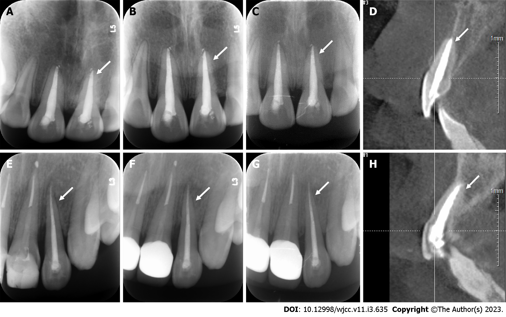
Figure 9 Periapical radiograph and cone beam computed tomography images of two cases during follow-up observation.
The periodontal membrane space was continuous, and no sign of pathological root absorption was observed in the avulsed teeth. A and E: 3-mo follow-up radiograph images of Cases 1 and 2; B and F: 6-mo follow-up radiograph images of Cases 1 and 2; C and G: 12-mo follow-up radiograph of Cases 1 and 2; D and H: 12-mo follow-up cone-beam computed tomography images of Cases 1 and 2.
- Citation: Yang Y, Liu YL, Jia LN, Wang JJ, Zhang M. Rescuing “hopeless” avulsed teeth using autologous platelet-rich fibrin following delayed reimplantation: Two case reports. World J Clin Cases 2023; 11(3): 635-644
- URL: https://www.wjgnet.com/2307-8960/full/v11/i3/635.htm
- DOI: https://dx.doi.org/10.12998/wjcc.v11.i3.635


