Copyright
©The Author(s) 2023.
World J Clin Cases. Jan 26, 2023; 11(3): 576-597
Published online Jan 26, 2023. doi: 10.12998/wjcc.v11.i3.576
Published online Jan 26, 2023. doi: 10.12998/wjcc.v11.i3.576
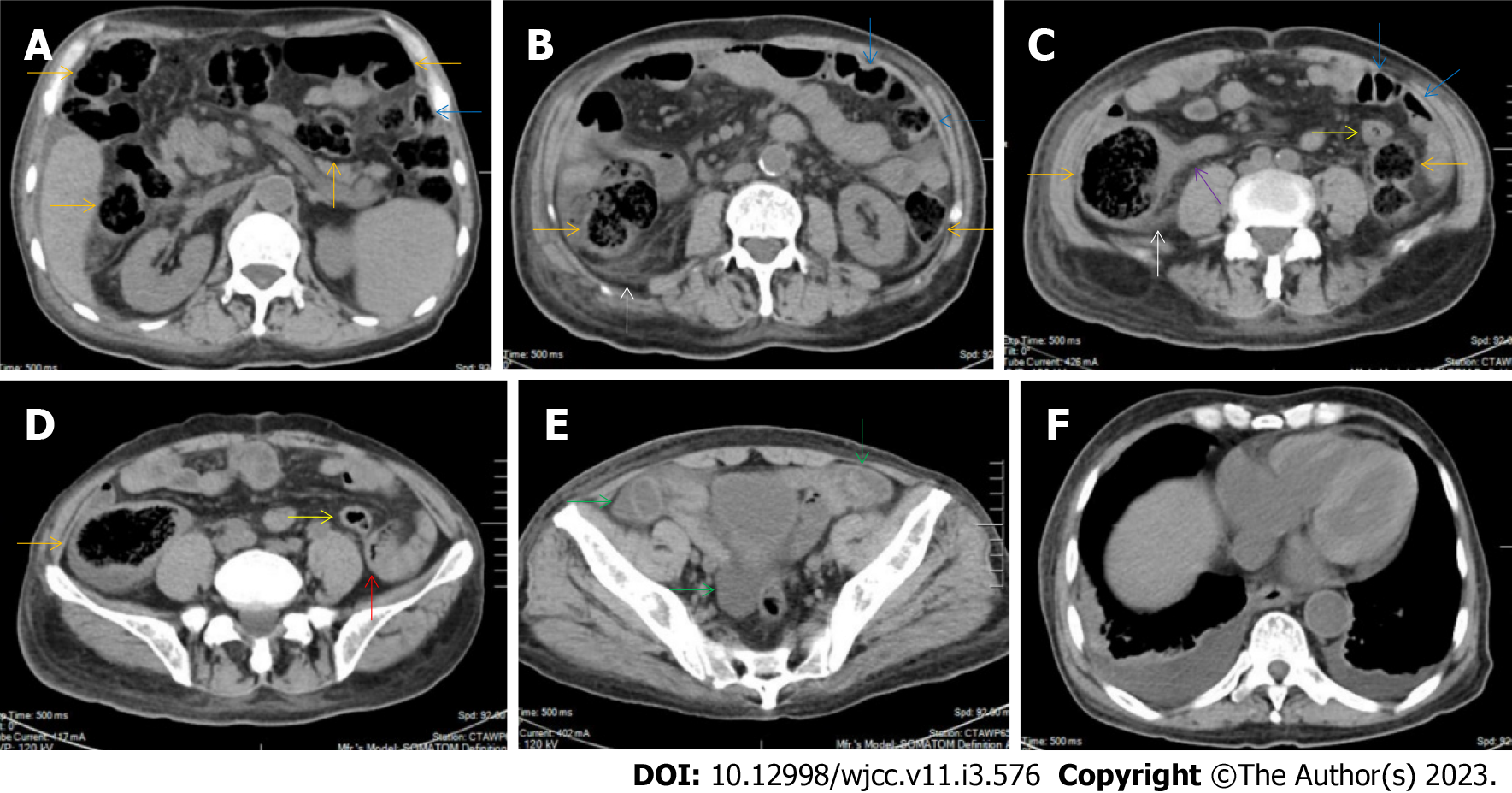
Figure 17 Characteristic images of case 5.
A-D: Characteristic images of the bowel inflammatory lesions. From the cecum to the descending colon, the lumen was dilated, the mucosa was hyperdence and the wall was thickened and stratified in some segments (orange arrows), with striking mesenteric fat stranding. There was a hypertrophic lesion (a red arrow) in the terminal descending colon. The mucosa of the proximal sigmoid colon was hyperdense and the lumen was gas-filled (blue arrows), following which was a short segment of strictured sigmoid colon (yellow arrows). The ileocecal valve and the terminal ileum were thickened and strictured but without mural stratification (a purple arrow), proximal to which the small intestine was liquid-filled. In the ileocecal region, the colonic wall was thickened with smudgy peritoneal thickening (white arrows); E: Characteristic image of the pelvic liquid collection. Mild ascites was present in both the right and left iliac fossa (green arrows), together with a thickened peritoneum suggesting the presence of peritoneal involvement; F: Characteristic image of the chest computed tomography (CT) scan. Chest CT showed the presence of pleural effusion in the bilateral cavities and bilateral pleural hypertrophic thickening. Enlarged blood vessels extended to the hypertrophic lesions.
- Citation: Zhao XC, Xue CJ, Song H, Gao BH, Han FS, Xiao SX. Bowel inflammatory presentations on computed tomography in adult patients with severe aplastic anemia during flared inflammatory episodes. World J Clin Cases 2023; 11(3): 576-597
- URL: https://www.wjgnet.com/2307-8960/full/v11/i3/576.htm
- DOI: https://dx.doi.org/10.12998/wjcc.v11.i3.576


