Copyright
©The Author(s) 2023.
World J Clin Cases. Jan 26, 2023; 11(3): 576-597
Published online Jan 26, 2023. doi: 10.12998/wjcc.v11.i3.576
Published online Jan 26, 2023. doi: 10.12998/wjcc.v11.i3.576
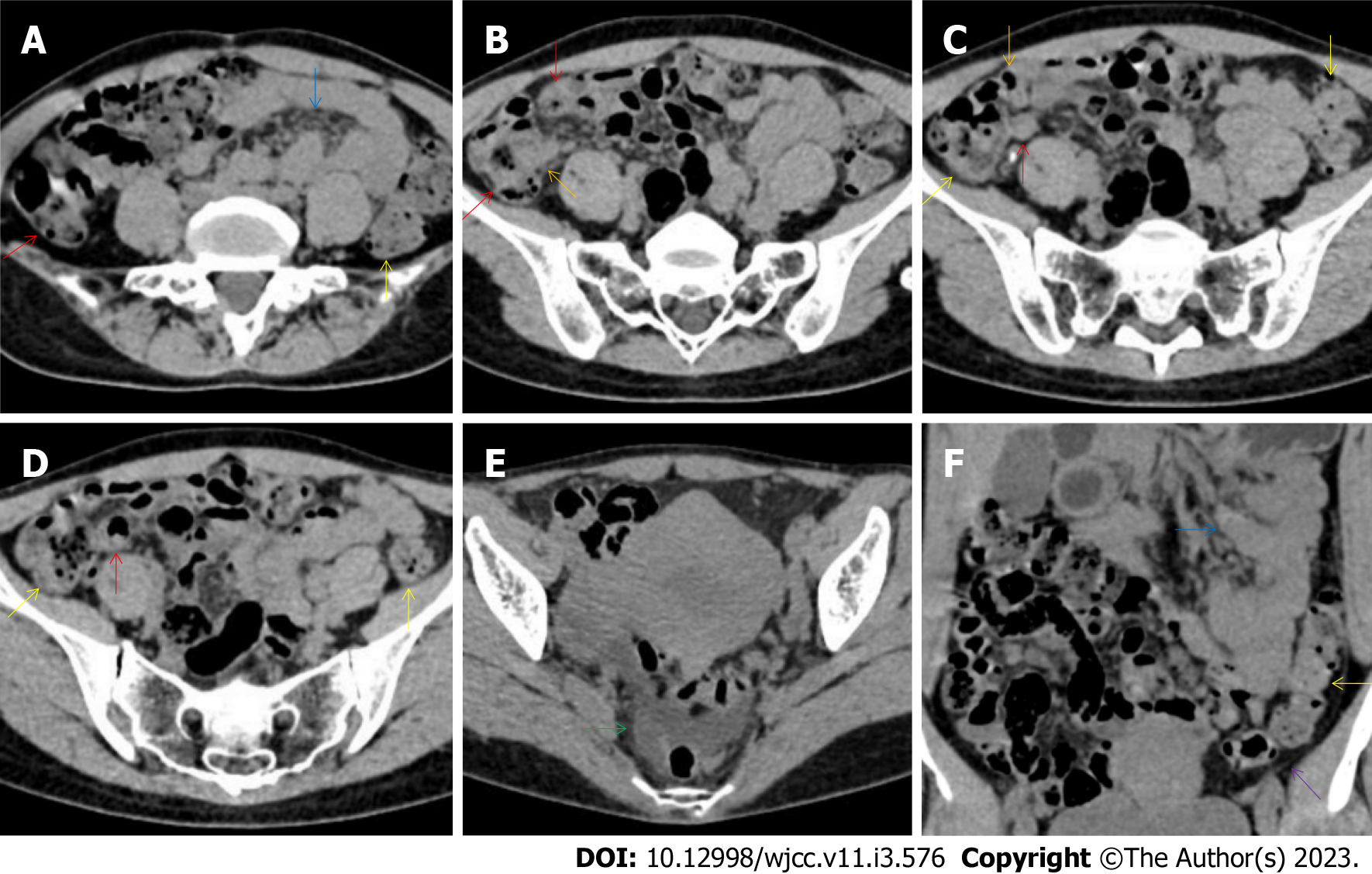
Figure 16 Characteristic images of case 12.
A-D: Characteristic images of the bowel inflammatory lesions. The ileocecal valve and the terminal ileal wall (red arrows) were thickened, stratified and strictured. Proximal to the strictured terminal ileum, the ileal lumen was dilated and gas-filled, and the mucosa was hyperdense. A large cluster of hypervasular mesenteric fat proliferation wrapped the dilated and gas-filled ileum. The jejunum was liquid-filled and the jejunual loop was adhesive. A cluster of hypervascular fat stranding wrapped a short segment of the jejunum (blue arrows), suggesting that the transmural inflammation was more serious in this jejunal segment. The colonic wall was also thickened and stratified, with intramural gas and subserosal pneumatosis (yellow arrows); E: Characteristic image of the pelvic liquid collection. Mild liquid collection was present in the pelvic cavity (a green arrow), together with the thickened peritoneum (a purple arrow) suggesting the presence of peritoneal involvement; F: Characteristic image in coronally reconstructed section. A coronally reconstructed image better outlined the above-mentioned imaging features.
- Citation: Zhao XC, Xue CJ, Song H, Gao BH, Han FS, Xiao SX. Bowel inflammatory presentations on computed tomography in adult patients with severe aplastic anemia during flared inflammatory episodes. World J Clin Cases 2023; 11(3): 576-597
- URL: https://www.wjgnet.com/2307-8960/full/v11/i3/576.htm
- DOI: https://dx.doi.org/10.12998/wjcc.v11.i3.576


